

The role of iron in the pathogenesis of endometriosis: a systematic review
- PMID: 37638130
- PMCID: PMC10457727
- DOI: 10.1093/hropen/hoad033
The role of iron in the pathogenesis of endometriosis: a systematic review
Abstract
Study question: What is the role of iron in the pathophysiology of endometriosis?
Summary answer: Iron excess is demonstrated wherever endometriotic tissues are found and is associated with oxidative stress, an inflammatory micro-environment, and cell damage; the iron-mediated oxidative stress is independently linked to subfertility, symptom severity, and malignant transformation.
What is known already: Iron is found in excess in endometriotic tissues, and multiple mechanisms have been studied and posited to explain this. It is clear that iron excess plays a vital role in promoting oxidative stress and cell damage. The evidence base is large, but no comprehensive reviews exist to summarize our understanding and highlight the overarching themes to further our understanding and suggest future directions of study for the field.
Study design size duration: This systematic review with a thematic analysis retrieved studies from the PubMed, Embase, Web of Science, and Cochrane Library databases and searches were conducted from inception through to August 2022. Human and animal studies published in the English language were included and identified using a combination of exploded MeSH terms ('Iron' and 'Endometriosis') and free-text search terms ('Iron', 'Ferric', 'Ferrous', 'Endometriosis', 'Endometrioma').
Participants/materials setting methods: This review was reported in accordance with the PRISMA guidelines. All studies reporting original data concerning the role of iron or iron complexes in the pathophysiology of endometriosis were included. Studies that did not report original data or provided a review of the field were excluded. Bias analysis was completed for each included study by using the Newcastle-Ottawa scoring system.
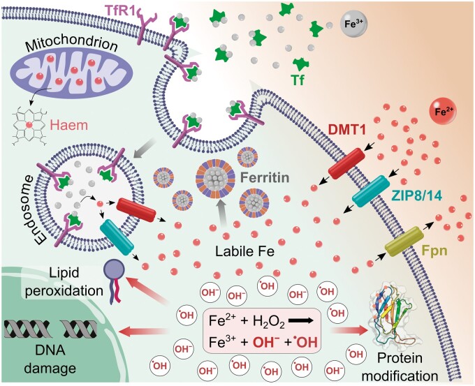
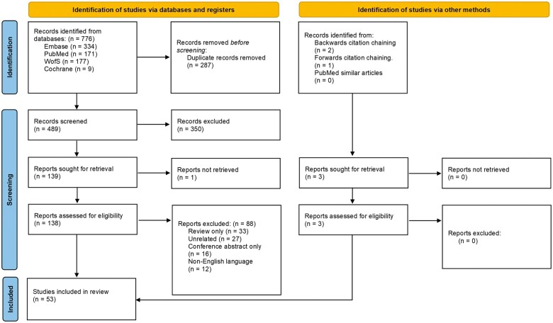
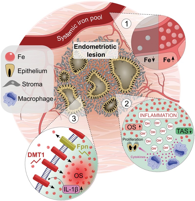
Main results and the role of chance: There were 776 records identified and these were screened down to 53 studies which met the eligibility criteria, including 6 animal and 47 human studies, with 3556 individual participants. Iron excess is demonstrated in various tissues and fluids, including ovarian endometriomas, ovarian follicles, ectopic endometriotic lesions, and peritoneal fluid. Markers of oxidative stress are strongly associated with high iron levels, and aberrant expression of iron-transport proteins has been demonstrated. Abnormal resistance to ferroptosis is likely. Iron-mediated oxidative stress is responsible for a pro-inflammatory micro-environment and is linked to subfertility, symptom severity, and, possibly, malignant transformation.
Limitations reasons for caution: A minority of the included studies were of objectively low quality with a high risk of bias and may lead to misleading conclusions. Additionally, multiple studies failed to appropriately characterize the included patients by known confounding variables, such as menstrual cycle phase, which may introduce bias to the findings.
Wider implications of the findings: Current literature depicts a central role of aberrant iron mechanics and subsequent oxidative stress in endometriosis. It is likely that iron excess is at least partly responsible for the persistence and proliferation of ectopic endometriotic lesions. As such, iron mechanics represent an attractive target for novel therapeutics, including iron chelators or effectors of the iron-oxidative stress pathway. There are significant gaps in our current understanding, and this review highlights and recommends several topics for further research. These include the role of iron chelation, resistance to ferroptosis, the relationship between iron excess and localized hypoxia, systemic iron pathophysiology in endometriosis, and the role of oxidative stress in malignant transformation.
Study funding/competing interests: J.W. and S.G.P. are supported by clinical fellowships at Liverpool University Hospital NHS Foundation trust. No additional funding was requested or required for the completion of this work. C.J.H. is supported by a Wellbeing of Women project grant (RG2137). D.K.H. is supported by a Wellbeing of Women project grant (RG2137) and an MRC clinical research training fellowship (MR/V007238/1). The authors have no conflicts of interest to declare.
Registration number: A protocol was prospectively registered with the PROSPERO database in August 2021 (CRD42021272818).
Keywords: endometriosis; ferroptosis; iron; iron chelation; iron excess; oxidative stress; systematic review.
© The Author(s) 2023. Published by Oxford University Press on behalf of European Society of Human Reproduction and Embryology.
Conflict of interest statement
The authors have no conflicts of interest to declare.
Figures
References
-
- Aisen P, Wessling-Resnick M, Leibold EA.. Iron metabolism. Curr Opin Chem Biol 1999;3:200–206. - PubMed
-
- Al-Shammaa NM. Effect of Iron relation with antioxidants on Endometriosis in red meat consumers females. Ann Trop Med Public Health 2020;23:887–893.
-
- Allavena G, Carrarelli P, DEL Bello B, Luisi S, Petraglia F, Maellaro E.. Autophagy is upregulated in ovarian endometriosis: a possible interplay with p53 and heme oxygenase-1. Fertil Steril 2015;103:1244–1251.e1. - PubMed
LinkOut - more resources
Full Text Sources
Medical
Research Materials
Miscellaneous