. 2023 Jul 7;5(1):vdad082.
doi: 10.1093/noajnl/vdad082. eCollection 2023 Jan-Dec.

Spectral flow cytometry identifies distinct nonneoplastic plasma extracellular vesicle phenotype in glioblastoma patients

Affiliations

Spectral flow cytometry identifies distinct nonneoplastic plasma extracellular vesicle phenotype in glioblastoma patients

Abudumijiti Zack Aibaidula et al. Neurooncol Adv. .

Abstract

Background: Glioblastoma (GBM) is the most common malignant brain tumor and has a poor prognosis. Imaging findings at diagnosis and in response to treatment are nonspecific. Developing noninvasive assays to augment imaging would be helpful. Plasma extracellular vesicles (EVs) are a promising biomarker source for this. Here, we develop spectral flow cytometry techniques that demonstrate differences in bulk plasma EV phenotype between GBM patients and normal donors that could serve as the basis of a liquid biopsy.

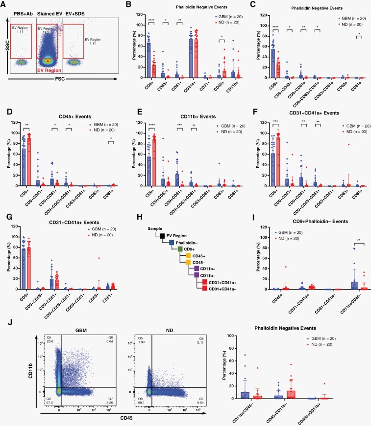
Methods: Plasma EVs were stained for EV-associated tetraspanins (CD9/CD63/CD81), markers indicating cell of origin (CD11b/CD31/CD41a/CD45), and actin/phalloidin (to exclude cell debris). EVs were analyzed using spectral flow cytometry. Multiparametric analysis using t-distributed stochastic neighbor embedding (t-SNE) and self-organizing maps on flow cytometry data (FlowSOM) was performed comparing GBM and normal donor (ND) plasma EVs.

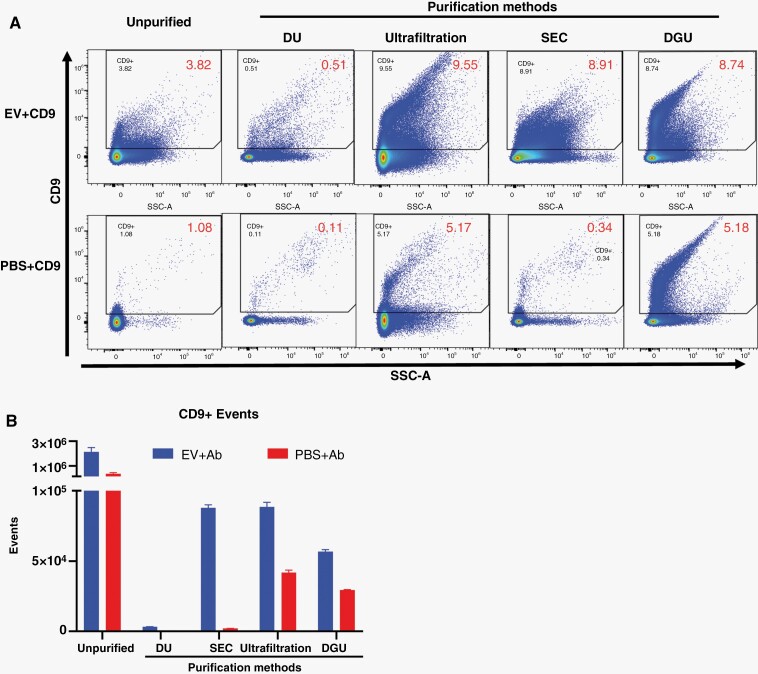
Results: Size exclusion chromatography plus spectral-based flow cytometer threshold settings enriched plasma EVs while minimizing background noise. GBM patients had increased CD9+, CD63+, CD81+, and myeloid-derived (CD11b+) EVs. Multiparametric analysis demonstrated distinct surface marker expression profiles in GBM plasma EVs compared to ND EVs. Fifteen plasma EV sub-populations differing in size and surface marker expression were identified, six enriched in GBM patients and two in normal donors.

Conclusions: Multiparametric analysis demonstrates that GBM patients have a distinct nonneoplastic plasma EV phenotype compared to ND. This simple rapid analysis can be performed without purifying tumor EVs and may serve as the basis of a liquid biopsy.

Keywords: extracellular vesicles; flow cytometry; glioblastoma; liquid biopsy.


Conflict of interest statement

no authors have relevant conflicts of interest to disclose.

Figures

Figure 1
Figure 1
Human plasma processing and EV staining assay controls. (A) Schematic showing method for plasma isolation from whole blood. (B) Settings of Cytek Aurora Flow cytometer were optimized to visualize different sizes of fluorescent and non-fluorescent ApogeeMix beads. (C) Serial dilution demonstrates a linear relationship between dilution and detected events for fluorescent beads (110 nm) and non-fluorescent beads (880nm), suggesting D, E clumping is absent. (D) EVs isolated from CD14+ monocytes in vitro were stained with CD9 (upper panel), CD11b (lower panel), and (E) CD9/CD11b together. Controls included buffer-only (PBS), buffer with reagent (PBS+Ab), single color-stained EVs (EV+Ab) and stained but detergent-treated EV samples (EV+Ab+SDS) (D, E). Serial dilution of CD9-stained EVs also demonstrated a linear relationship between dilution and detected events.
Figure 2
Figure 2
Comparing methods to separate and concentrate stained EVs from unbound antibodies. (A) Representative dot plots for CD9-stained EVs from CD14+ monocytes in vitro without removing unbound antibodies compared to after differential ultracentrifugation (DU), ultrafiltration, size exclusion chromatography (SEC), or density gradient ultracentrifugation (DGU) to eliminate excess antibodies. PBS+ CD9 antibodies were used as process control (lower panel). (B) All four methods reduced EV yield, but SEC was most effective at separating/concentrating EVs with the highest signal-to-noise ratio.
Figure 3
Figure 3
EV staining panel, assay controls, and data acquisition for plasma EV analysis. (A) Schematic outlining final staining and separation/concentration methods for plasma EVs. (B, C) CD9 antibody titration for plasma EVs samples did not show significant differences in event rates or median fluorescent intensity (MFI) with increasing amounts of CD9 antibodies. (D) Representative dot plots showing plasma EV surface marker expression determined using an SSC threshold for EV-associated tetraspanins (CD9, CD63, CD81), phalloidin (staining actin as a marker of contaminating cell debris), and leukocyte/platelet/endothelial markers (CD11b, CD31, CD41a, CD45). Assays controls included buffer only (PBS), buffer with reagents (PBS + all antibodies), single color staining, all color staining, and fluorescence minus one (FMO; all antibodies except one directed against the marker of interest). (E). Changing from a size-based SSC threshold for event acquisition (upper panel) to a spectral fluorescence-based surface marker expression threshold (lower panel) markedly reduced non-specific background staining as best demonstrated for CD9 expression in SEC-purified stained EVs. ns = not significant.
Figure 4
Figure 4
Analysis of plasma EVs in GBM and normal donors. (A) Dot plots showing gating strategy and controls for particles in the EV size range (EV region). (B) Pooled data from 20 GBM patients and 20 normal donors (ND) showing percentage of EV-associated tetraspanin (CD9, CD63, CD81) and cell of origin (CD45, CD11b, CD31, CD41a) surface marker expression among EV region/phalloidin-negative particles. CD9+ and CD41a+ EVs were most abundant. Tetraspanin expression was more common in GBM patients than ND. (C) Most plasma EVs expressed CD9 tetraspanin only. GBM patients had increased CD9+, CD9+/CD81+, CD9+/CD63+/CD81+ EVs. (D-G). Most CD45+, CD11b+, CD31+/CD41a+ and CD31+/CD41a- EV populations in ND only expressed the tetraspanin CD9 while GBM patients had increased frequencies of CD9+, CD9+/CD81+ and CD9+/CD63+/CD81+ EVs. (H) Strategy for tracing cell of origin for CD9+/Phalloidin- EVs. (I) No significant differences between GBM and ND CD45+ (leukocyte common antigen) and CD31+CD41a- (endothelial) plasma EVs. GBM patients had increased CD11b+/CD45- EVs (presumed myeloid origin) while ND had increased CD31+CD41a+ (platelet-derived) EVs. (J) Strikingly, most CD11b+ plasma EVs do not express CD45. This is distinct from myeloid cells which would typically be CD11b+/CD45+. *p <0.05, **p < 0.01, ***p < 0.001, ****p < 0.0001.
Figure 5
Figure 5
T-SNE and FlowSOM analysis reveal differently expressed plasma EV subpopulations in GBM and normal donors. (A) T-SNE (t-distributed stochastic neighbor embedding) analysis based on SSC, CD9, CD81, CD63, CD31, CD45, CD11b, and CD41a reveals different plasma EV clustering features for GBM patients and normal donors. (B) FlowSOM (self-organizing maps of flow cytometry data) analysis reveals 15 EV subpopulations differing in size (SSC) and surface marker expression. (C) Some groups are enriched in ND (Pop0, Pop2) while others are enriched in GBM (Pop4, Pop6, Pop7, Pop8, Pop10, Pop11). *p < 0.05, ***p < 0.001, ****p < 0.0001.

References

    1. Ostrom QT, Cioffi G, Gittleman H, et al. . CBTRUS statistical report: primary brain and other central nervous system tumors diagnosed in the United States in 2012-2016. Neuro Oncol. 2019;21(Suppl 5):v1–v100. - PMC - PubMed
    1. Ostrom QT, Gittleman H, Stetson L, Virk S, Barnholtz-Sloan JS. Epidemiology of intracranial gliomas. Prog Neurol Surg. 2018;30:1–11. - PubMed
    1. Nam JY, de Groot JF.. Treatment of glioblastoma. J Oncol Pract. 2017;13(10):629–638. - PubMed
    1. Marenco-Hillembrand L, Wijesekera O, Suarez-Meade P, et al. . Trends in glioblastoma: outcomes over time and type of intervention: a systematic evidence based analysis. J Neurooncol. 2020;147(2):297–307. - PubMed
    1. Stupp R, Mason WP, van den Bent MJ, et al. ; European Organisation for Research and Treatment of Cancer Brain Tumor and Radiotherapy Groups. Radiotherapy plus concomitant and adjuvant temozolomide for glioblastoma. N Engl J Med. 2005;352(10):987–996. - PubMed

LinkOut - more resources