Review
. 2024 Jan 1;31(1):1-14.
doi: 10.1097/PAP.0000000000000411. Epub 2023 Aug 28.

Invasive Squamous Cell Carcinoma of the Cervix: A Review of Morphological Appearances Encountered in Human Papillomavirus-associated and Papillomavirus-independent Tumors and Precursor Lesions

Affiliations
Review

Invasive Squamous Cell Carcinoma of the Cervix: A Review of Morphological Appearances Encountered in Human Papillomavirus-associated and Papillomavirus-independent Tumors and Precursor Lesions

Simona Stolnicu et al. Adv Anat Pathol. .

Abstract

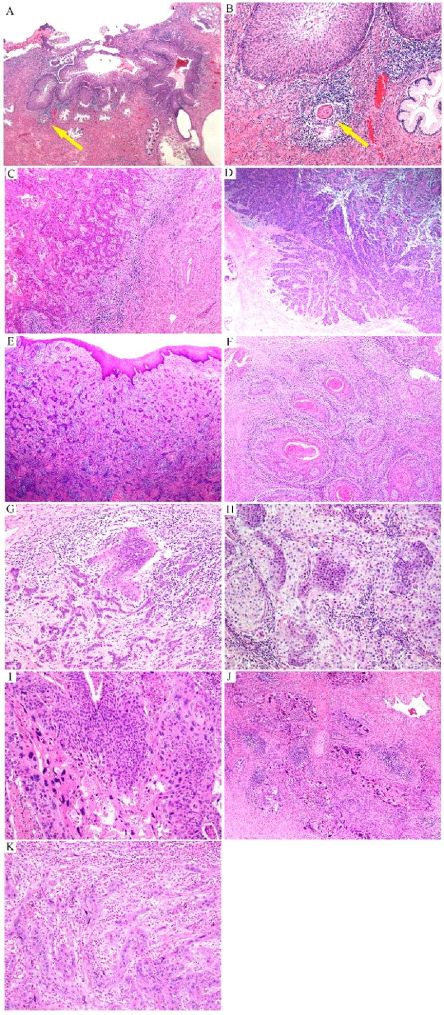
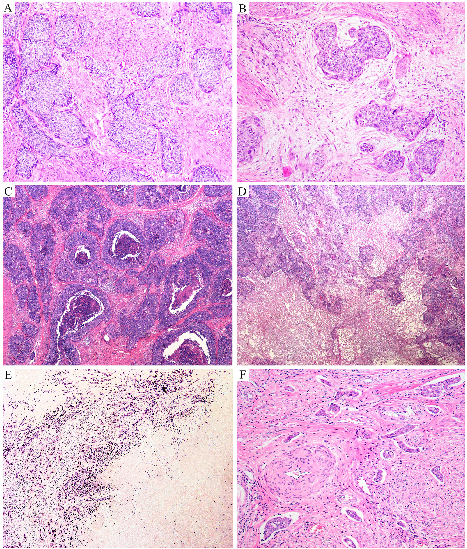
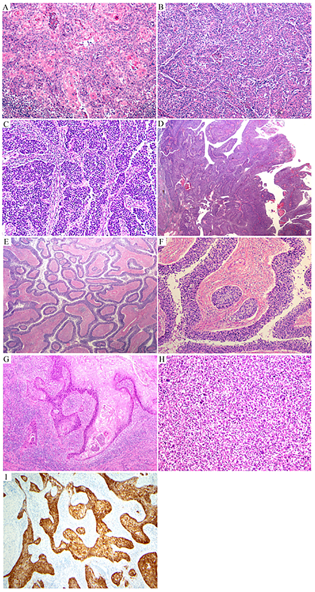
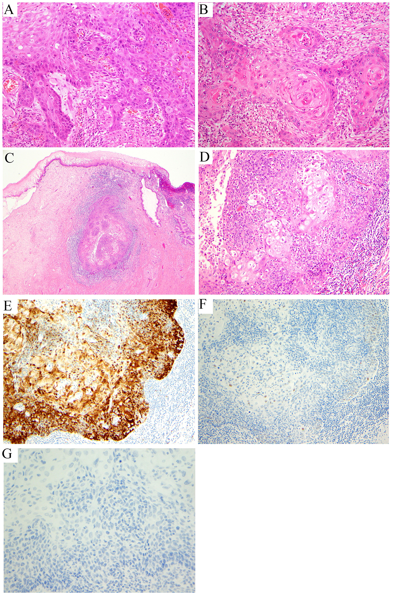
Cervical cancer is the fourth most common cancer among women globally. Historically, human papillomavirus (HPV) infection was considered necessary for the development of both precursor and invasive epithelial tumors of the cervix; however, studies in the last decade have shown that a significant proportion of cervical carcinomas are HPV-independent (HPVI). The 2020 World Health Organization (WHO) Classification of Female Genital Tumors separates both squamous cell carcinomas (SCCs) and endocervical adenocarcinomas (ECAs) by HPV status into HPV-associated (HPVA) and HPVI tumors. The classification further indicates that, in contrast to endocervical adenocarcinomas, HPVI and HPVA SCCs cannot be distinguished by morphological criteria alone and suggests that HPV testing or correlates thereof are required for correct classification. Moreover, while HPVA SCC precursor lesions (ie, high-grade squamous intraepithelial lesion) are well known and characterized, precursors to HPVI SCCs have only been described recently in a small number of cases. We studied 670 cases of SCCs from the International Squamous Cell Carcinoma Project (ISCCP) to analyze the reproducibility of recognition of invasive SCC growth patterns, presence of lymphovascular space invasion, tumor grade, and associations with patient outcomes. Consistent with previous studies, we found histologic growth patterns and tumor types had limited prognostic implications. In addition, we describe the wide morphologic spectrum of HPVA and HPVI SCCs and their precursor lesions, including tumor growth patterns, particular and peculiar morphologic features that can lead to differential diagnoses, and the role of ancillary studies in the diagnosis of these tumors.


Conflict of interest statement

Outside the current work, A.I. reports consulting fees from Mylan. The remaining authors have no conflicts of interest to disclose.

Figures

Figure 1:
Figure 1:. Infiltrating human papillomavirus-associated (HPVA) squamous cell carcinoma (SCC):
extensive high-grade squamous intraepithelial lesion (HSIL) with a tiny focus of invasion (arrow) (A) characterized by a small nest of tumor cells with paradoxical maturation (arrow) (B); invasive SCC with ragged epithelial-stromal interface (C); tumor cell buds invade the stroma with associated stromal desmoplasia (D, E); invasive buds of various sizes and shapes with central keratin pearls (F); buds and cords of tumor cells infiltrate the stroma in association with inflammatory infiltrates (G); infiltrating tumor cells display paradoxical maturation and uniform, small nuclei (H) or highly atypical nuclei (I, J) sometimes with a spindle shape, mimicking a sarcoma (K).
Figure 2:
Figure 2:. Infiltrating human papillomavirus-associated (HPVA) squamous cell carcinoma (SCC):
fibrous stroma (A); myxoid stroma (B); areas of comedo-type of necrosis (C) or massive necrosis (D, E); numerous foci of lymphovascular space invasion (LVSI) at the periphery of the tumor (F).
Figure 3:
Figure 3:. Microscopic growth patterns in human papillomavirus-associated (HPVA) squamous cell carcinomas (SCCs):
keratinizing (A); non-keratinizing (B); basaloid (C); papillary (D); inverted papillary (papillae invading the storm without an exophytic component) (E, F); warty (G); lymphoepithelioma-like (H); microscopic growth pattern, p16 is block-type positive (I).
Figure 4:
Figure 4:. Peculiar architectural patterns in human papillomavirus-associated (HPVA) squamous cell carcinomas (SCCs):
palisading (A, B); pseudoglandular pattern (C); papillary SCC with pseudoglandular areas (D, E); Silva pattern A (F); Silva pattern B (G, H); Silva pattern C (I); inverted papillary pattern (J); adenoid basal-like morphology (K); adenoid cystic-like morphology (L).
Figure 5:
Figure 5:. Peculiar architectural patterns in human papillomavirus-associated (HPVA) squamous cell carcinomas (SCCs):
lobular architecture (A); micropapillary architecture (B); massive squamous benign metaplasia at the top of the tumor (C) associated with numerous calcifications (D); giant multinucleate cells associated with nests of tumor cells (E); massive calcification (F); psammoma bodies (G).
Figure 6:
Figure 6:. Peculiar cytologic features in human papillomavirus-associated (HPVA) squamous cell carcinomas (SCCs):
cytoplasmic clearing (A, B); rare cytoplasmic mucin (C, D); light and dark pattern (E); eosinophilic single cells (F); glassy-like morphology (G); multinucleated cells (H); dark and light pattern (I, J); psammoma bodies (K); sarcomatoid features (L).
Figure 7:
Figure 7:. Human papillomavirus-associated (HPVA) precursor lesions:
high-grade squamous intraepithelial lesion (HSIL), cervical intraepithelial neoplasia (CIN) grade 2) (A, B); HSIL, CIN grade 3 (C).
Figure 8:
Figure 8:. Human papillomavirus-independent (HPVI) precursor and infiltrating squamous cell carcinoma (SCC):
high-grade squamous intraepithelial lesion (HPVI) precursor lesion differentiated vulvar intraepithelial neoplasia (dVIN)-like (A); invasive HPVI keratinizing SCC with a dense inflammatory infiltrate (B); invasive HPVI SCC with a non-keratinizing growth pattern (C, D), p16 patchy (not overexpressed) (E), p53 wild type (patchy, low intensity nuclear staining) (F), HPV in situ hybridization (ISH) negative (G).

Similar articles

Cited by

References

    1. WHO Classification of Tumours Female Genital Tumours. 5 ed. WHO Classification of Tumours. International Agency for Research on Cancer; 2020.
    1. Siegel RL, Miller KD, Fuchs HE, Jemal A. Cancer statistics, 2022. CA Cancer J Clin. Jan 2022;72(1):7–33. doi:10.3322/caac.21708 - DOI - PubMed
    1. Burd EM. Human papillomavirus and cervical cancer. Clin Microbiol Rev. Jan 2003;16(1):1–17. doi:10.1128/cmr.16.1.1-17.2003 - DOI - PMC - PubMed
    1. Kurman RJ, Carcangiu ML, Herrington CS. World Health Organisation classification of tumours of the female reproductive organs. 4 ed. World Health Organization classification of tumours Lyon: International agency for research on cancer; 2014.
    1. Park KJ, Soslow RA. Current concepts in cervical pathology. Arch Pathol Lab Med. May 2009;133(5):729–38. doi:10.5858/133.5.729 - DOI - PubMed