. 2023 Sep 14;91(9):e0015023.
doi: 10.1128/iai.00150-23. Epub 2023 Aug 28.

Role of the CagY antenna projection in Helicobacter pylori Cag type IV secretion system activity

Affiliations

Role of the CagY antenna projection in Helicobacter pylori Cag type IV secretion system activity

Sirena C Tran et al. Infect Immun. .

Abstract

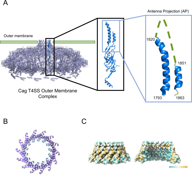
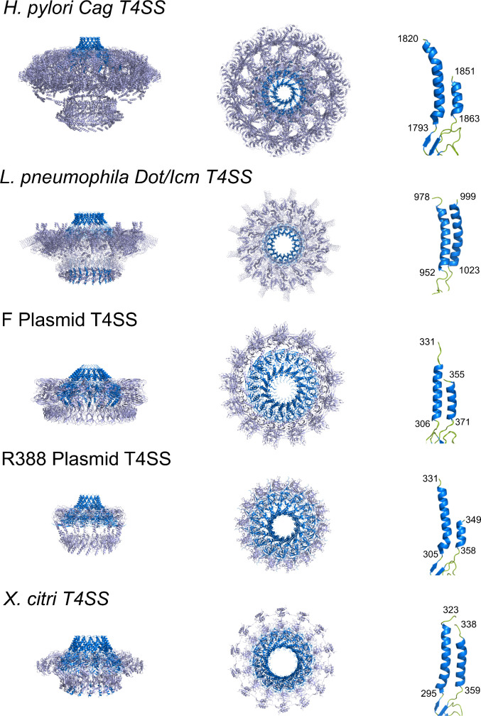
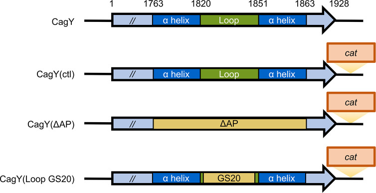
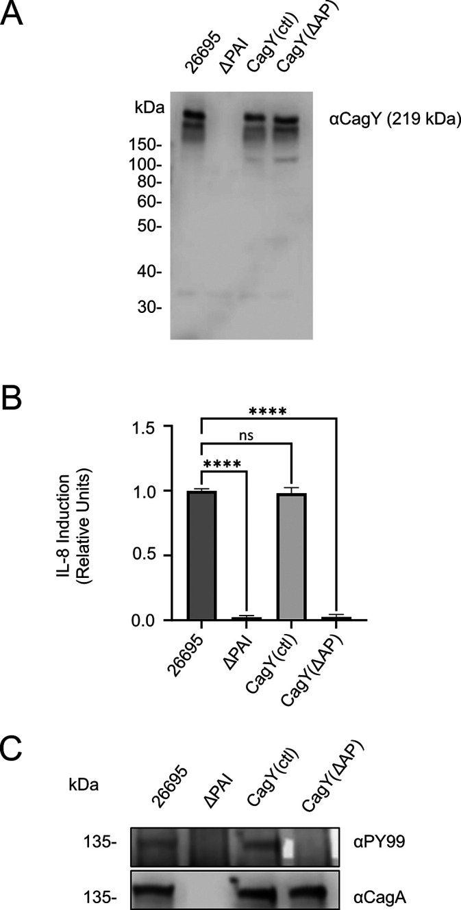
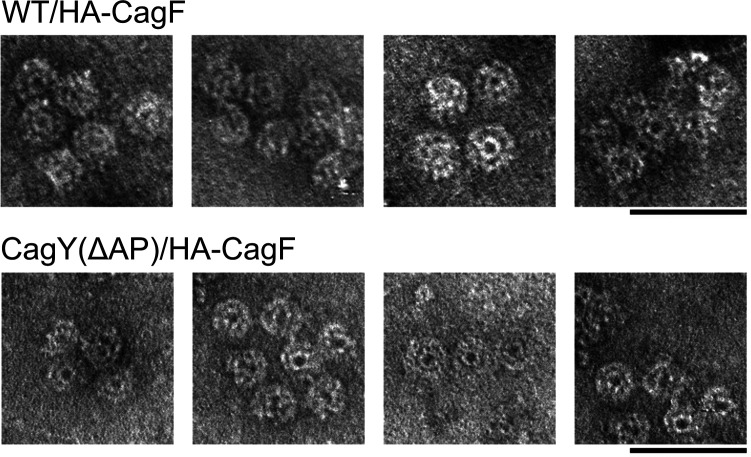
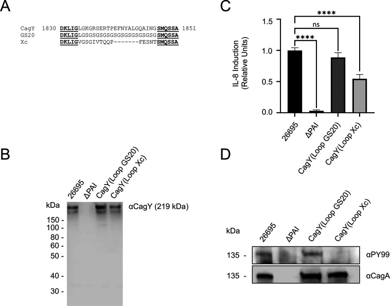
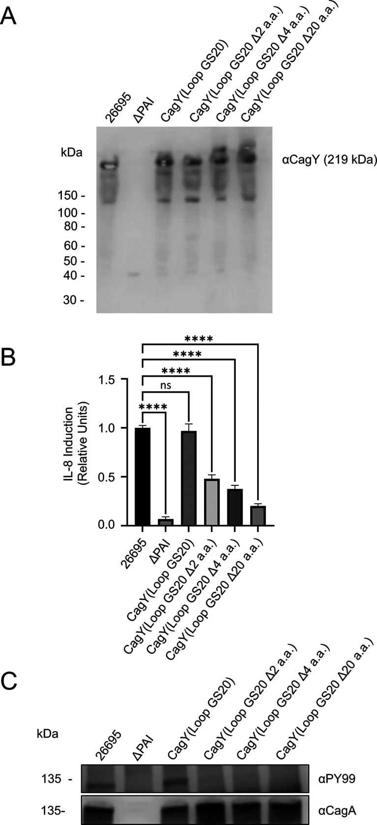
Helicobacter pylori strains containing the cag pathogenicity island (PAI) are associated with the development of gastric adenocarcinoma and peptic ulcer disease. The cag PAI encodes a secreted effector protein (CagA) and a type IV secretion system (Cag T4SS). Cag T4SS activity is required for the delivery of CagA and non-protein substrates into host cells. The Cag T4SS outer membrane core complex (OMCC) contains a channel-like domain formed by helix-loop-helix elements (antenna projections, AP) from 14 copies of the CagY protein (a VirB10 ortholog). Similar VirB10 antenna regions are present in T4SS OMCCs from multiple bacterial species and are predicted to span the outer membrane. In this study, we investigated the role of the CagY antenna region in Cag T4SS OMCC assembly and Cag T4SS function. An H. pylori mutant strain with deletion of the entire CagY AP (∆AP) retained the capacity to produce CagY and assemble an OMCC, but it lacked T4SS activity (CagA translocation and IL-8 induction in AGS gastric epithelial cells). In contrast, a mutant strain with Gly-Ser substitutions in the unstructured CagY AP loop retained Cag T4SS activity. Mutants containing CagY AP loops with shortened lengths were defective in CagA translocation and exhibited reduced IL-8-inducing activity compared to control strains. These data indicate that the CagY AP region is required for Cag T4SS activity and that Cag T4SS activity can be modulated by altering the length of the CagY AP unstructured loop.

Keywords: bacterial outer membrane protein; bacterial secretion system; gastric cancer; membrane channel; pathogenicity island.


Conflict of interest statement

The authors declare no conflict of interest.

Figures

Fig 1
Fig 1
Schematic representation of the CagY antenna projection. (A) A ribbon diagram of the outer membrane cap (OMC) portion of the Cag T4SS outer membrane core complex (OMCC). Left panel: side view of the OMCC shows the localization of a single CagY protein (blue color). Middle panel: structure of the portion of CagY localized to the OMC, which includes the antenna projection (AP). Right panel: enlargement shows that the AP is composed of two α-helices connected by an unstructured loop (green dashed line drawn in for reference). (B) A top-down ribbon diagram view of the CagY antenna region illustrates the antenna region that is formed by 14 CagY APs. (C) A hydrophobicity analysis of the CagY antenna region shows that the exterior surface is predicted to be hydrophobic, while the interior surface is hydrophilic. Blue indicates hydrophilicity, and yellow indicates hydrophobicity.
Fig 2
Fig 2
Conservation of VirB10 antenna regions in multiple T4SSs. The left and middle columns show side and top-down views of outer membrane core complexes from five different bacterial T4SSs. Both the OMC and PR are shown for the H. pylori and L. pneumophila T4SSs. Areas highlighted in blue are the VirB10 homologs. The right column shows images of VirB10 APs labeled with corresponding amino acid numbers. Green coloring indicates loops, connecting α helices or β strands, that were resolved in the structures. From top to bottom: H. pylori Cag T4SS (PDB: 6X6S and 6X6J), L. pneumophila Dot/Icm T4SS (PDB: 7MUY), F plasmid-encoded T4SS (PDB: 7SPC & 7SPB), R388 plasmid-encoded T4SS (PDB: 7O3J &7O3T), and Xanthomonas citri T4SS (PDB: 6GYB).
Fig 3
Fig 3
Schematic of CagY antenna projection (AP) mutations. The schematic depicts alterations to the AP region of CagY. CagY(ctl): Insertion of cat gene downstream of CagY (strain SCT13). CagY(∆AP): deletion of the entire antenna region (SCT20). CagY(Loop GS20): 20 amino acids of the unstructured loop of the AP were replaced with 10 glycine and serine (Gly-Ser) repeats (SCT70). cat, chloramphenicol acetyltransferase.
Fig 4
Fig 4
The CagY antenna projection is required for Cag T4SS activity. To generate a strain in which the portion of cagY encoding the AP was deleted [CagY(∆AP), also named SCT20], a cat gene downstream of cagY was used as a selectable marker. CagY(ctl) is a control strain (SCT13) containing the cat gene downstream of cagY, without alteration of cagY. The wild type (WT) 26695 strain was used as a positive control, and a cag ∆PAI mutant was used as a negative control. (A) Production of CagY detected by western blotting. Immunoreactive bands with molecular masses <219 kDa represent CagY degradation products. (B) H. pylori strains were co-cultured with AGS gastric cells, and Cag T4SS activity was assessed by an IL-8 induction assay. The IL-8 data represent three or more independent experiments, each with multiple technical replicates. The values represent the mean ± SEM. The significance of differences compared to strain 26695 was determined by a One-Way ANOVA and Dunnett’s multiple comparisons test. ****P < 0.0001. (C) CagA translocation assay using AGS cells, as described in the Methods section. The CagA translocation figure shows representative results from one of three independent experiments. αPY99, antiphosphotyrosine antibody. Collectively, the data show that the mutant strains produce CagY and that the AP is required for both IL-8 induction and CagA translocation.
Fig 5
Fig 5
The CagY antenna region is not required for core complex assembly. Cag T4SS outer membrane core complexes were isolated from both HA-CagF and CagY(∆AP) HA-CagF (strain SCT73) using an immunopurification protocol described in Methods. Samples were analyzed via negative stain electron microscopy to visualize the Cag T4SS OMCCs. Scale bar, 100 nm.
Fig 6
Fig 6
The specific sequence of the CagY antenna projection (AP) loop is not required for Cag T4SS activity. Mutants were generated to assess the functional properties of the CagY AP loop. In the CagY(Loop GS20) mutant (SCT70), 20 amino acids in the middle of the loop region were replaced with repeating glycine/serine residues. In the CagY(Loop Xc) mutant (SCT67), the middle 22 amino acids in the loop region were replaced with corresponding amino acids from the Xanthomonas citri VirB10 loop sequence. (A) A sequence alignment of wild-type CagY and CagY AP loop mutants. GS20: CagY(Loop GS20) mutant; Xc: CagY(Loop Xc) mutant. Sequences bolded and underlined show the CagY sequences that were not altered, while sequences in the middle show the altered sequences. (B) Western blot analysis indicated that the production of CagY was not disrupted in the CagY mutant strains. (C) H. pylori strains were co-cultured with AGS gastric epithelial cells, and Cag T4SS activity was assessed by an IL-8 induction assay. The IL-8 data represent the results of three or more independent experiments, each with multiple technical replicates. The values represent the mean ± SEM. The significance of differences compared to strain 26695 was determined by a One-Way ANOVA and Dunnett’s multiple comparisons test. ****P ≤ 0.0001. (D) Analysis of CagA translocation. The CagA translocation figure shows representative results from one of three independent experiments. αPY99, anti-phosphotyrosine antibody. Collectively, the data show the mutant strains produce CagY, and mutants with substitution mutations in the CagY loop do not have a defect in T4SS activity. However, CagA translocation is disrupted and IL-8 induction is reduced in the CagY Loop Xc mutant.
Fig 7
Fig 7
The length of the CagY AP loop influences Cag T4SS activity. The CagY antenna loop region of the CagY(Loop GS20) strain (SCT70) was altered by introducing variations in the length of the region containing Gly-Ser residues. (A) Western blot analysis shows that the shortening of the loop does not affect the production of CagY. Immunoreactive bands with molecular masses <219 kDa represent CagY degradation products. (B) H. pylori strains were co-cultured with AGS gastric epithelial cells, and Cag T4SS activity was assessed by an IL-8 induction assay. The IL-8 data represent the results of three or more independent experiments, each with multiple technical replicates. The values represent the mean ± SEM. The significance of differences compared to strain 26695 was determined by One-Way ANOVA and Dunnett’s multiple comparisons test. ****P ≤ 0.0001. (C) Analysis of CagA translocation. The CagA translocation figure shows representative results from one of the three independent experiments. αPY99, anti-phosphotyrosine antibody.

Similar articles

Cited by

References

    1. Hooi JKY, Lai WY, Ng WK, Suen MMY, Underwood FE, Tanyingoh D, Malfertheiner P, Graham DY, Wong VWS, Wu JCY, Chan FKL, Sung JJY, Kaplan GG, Ng SC. 2017. Global prevalence of Helicobacter pylori infection: systematic review and meta-analysis. Gastroenterology 153:420–429. doi:10.1053/j.gastro.2017.04.022 - DOI - PubMed
    1. Atherton JC, Blaser MJ. 2009. Coadaptation of Helicobacter pylori and humans: ancient history, modern implications. J Clin Invest 119:2475–2487. doi:10.1172/JCI38605 - DOI - PMC - PubMed
    1. Fox JG, Wang TC. 2007. Inflammation, atrophy, and gastric cancer. J Clin Invest 117:60–69. doi:10.1172/JCI30111 - DOI - PMC - PubMed
    1. Cover TL, Blaser MJ. 2009. Helicobacter pylori in health and disease. Gastroenterology 136:1863–1873. doi:10.1053/j.gastro.2009.01.073 - DOI - PMC - PubMed
    1. de Martel C, Georges D, Bray F, Ferlay J, Clifford GM. 2020. Global burden of cancer attributable to infections in 2018: a worldwide incidence analysis. Lancet Glob Health 8:e180–e190. doi:10.1016/S2214-109X(19)30488-7 - DOI - PubMed

Publication types

LinkOut - more resources