. 2023 Oct 13;51(18):9733-9747.
doi: 10.1093/nar/gkad686.

RAP80 phase separation at DNA double-strand break promotes BRCA1 recruitment

Affiliations

RAP80 phase separation at DNA double-strand break promotes BRCA1 recruitment

Caolitao Qin et al. Nucleic Acids Res. .

Abstract

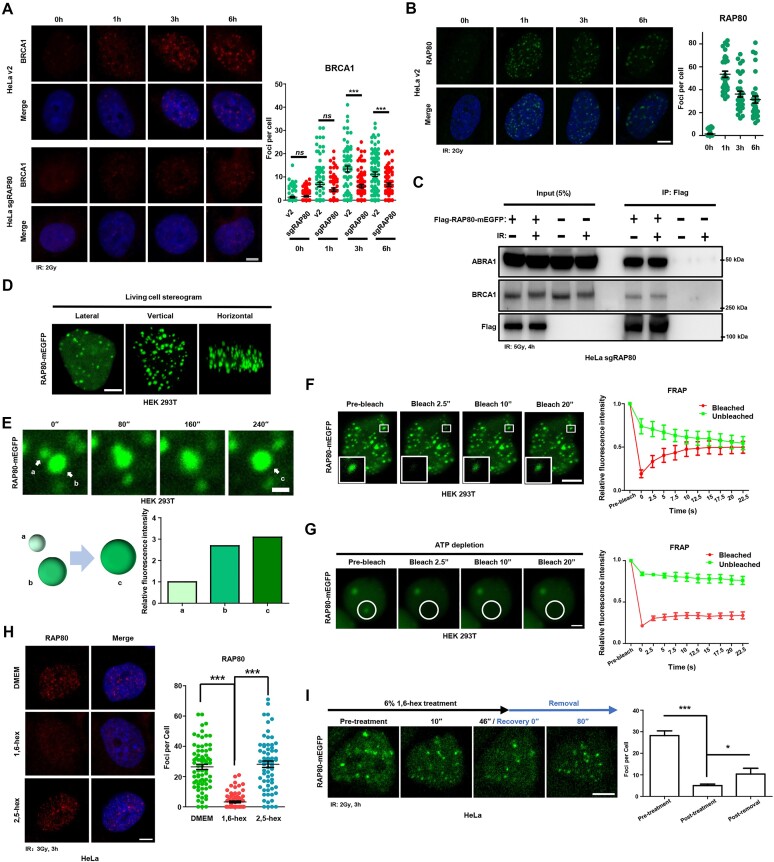
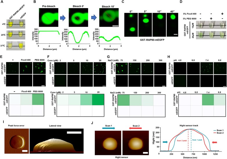
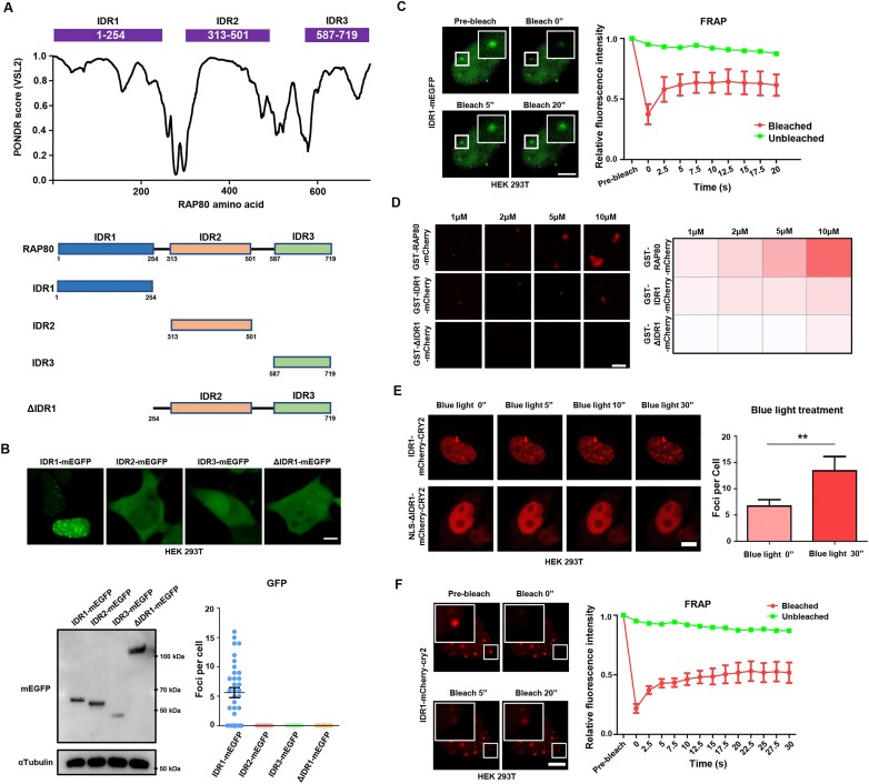
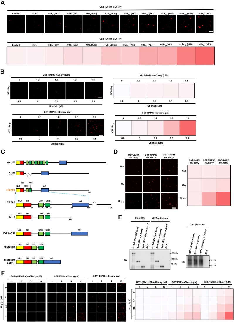
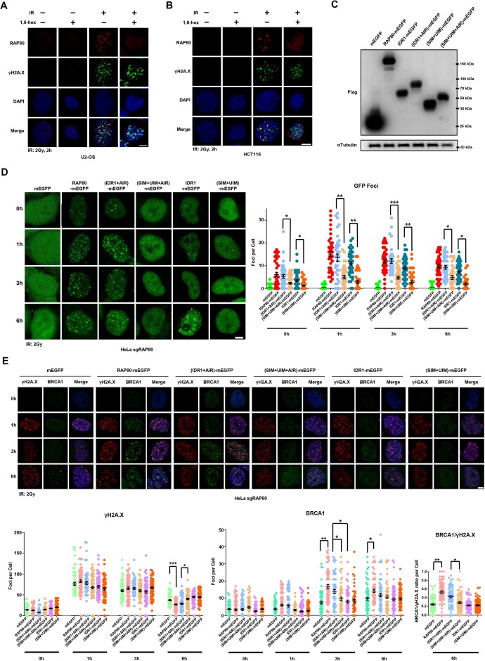
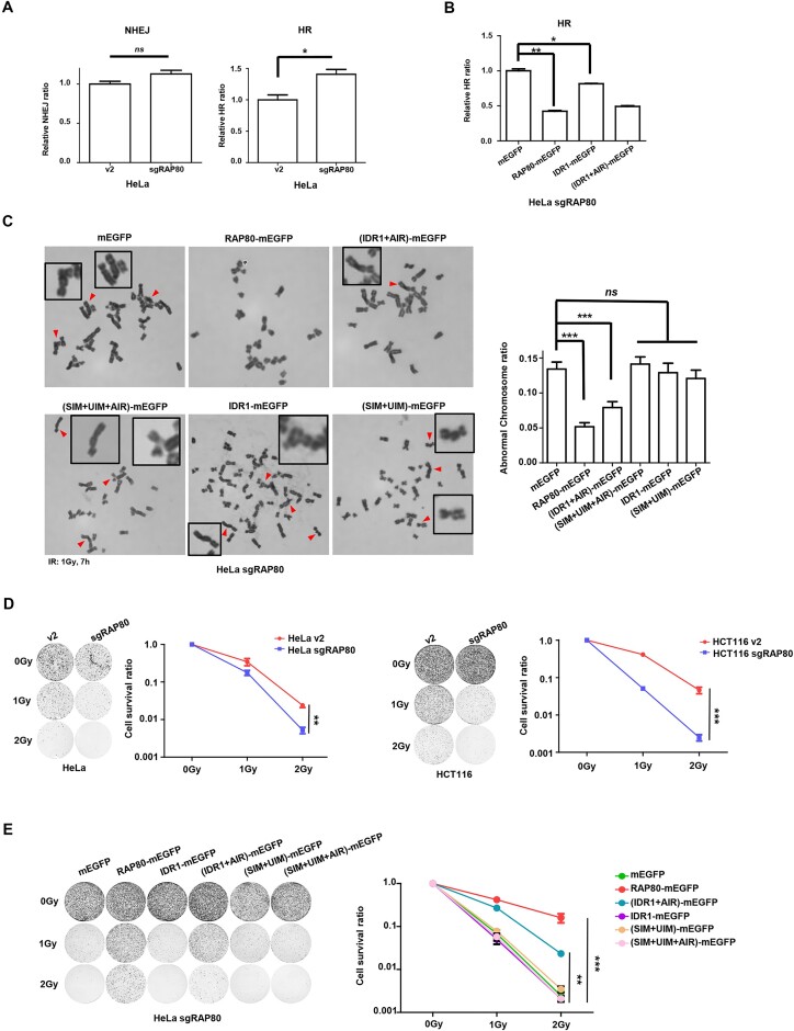
RAP80 has been characterized as a component of the BRCA1-A complex and is responsible for the recruitment of BRCA1 to DNA double-strand breaks (DSBs). However, we and others found that the recruitment of RAP80 and BRCA1 were not absolutely temporally synchronized, indicating that other mechanisms, apart from physical interaction, might be implicated. Recently, liquid-liquid phase separation (LLPS) has been characterized as a novel mechanism for the organization of key signaling molecules to drive their particular cellular functions. Here, we characterized that RAP80 LLPS at DSB was required for RAP80-mediated BRCA1 recruitment. Both cellular and in vitro experiments showed that RAP80 phase separated at DSB, which was ascribed to a highly disordered region (IDR) at its N-terminal. Meanwhile, the Lys63-linked poly-ubiquitin chains that quickly formed after DSBs occur, strongly enhanced RAP80 phase separation and were responsible for the induction of RAP80 condensation at the DSB site. Most importantly, abolishing the condensation of RAP80 significantly suppressed the formation of BRCA1 foci, encovering a pivotal role of RAP80 condensates in BRCA1 recruitment and radiosensitivity. Together, our study disclosed a new mechanism underlying RAP80-mediated BRCA1 recruitment, which provided new insight into the role of phase separation in DSB repair.


Figures

Graphical Abstract
Graphical Abstract
Figure 1.
Figure 1.
RAP80 forms liquid-like condensates in the nucleus. (A, B) HeLa cells were exposed to 2 Gy irradiation and fixed at the indicated time before IF analysis. Scale bar, 5 μm. (C) CoIP assay showed the association of Flag-RAP80-mEGFP, BRCA1 and ABRA1 in HeLa sgRAP80 cells. (D) Confocal image 3D reconstruction of RAP80-mEGFP droplets in the nucleus. Scale bar, 5 μm. (E) Confocal image sequence showing the fusion between adjacent RAP80-mEGFP foci in the nucleus. The quantification data showed the fluorescence intensity of droplet a, b and c. Scale bar 5 μm. (F) FRAP assay of RAP80-mEGFP puncta in HEK 293T cells. Scale bar, 5 μm. (G) ATP depletion abolished the FRAP of RAP80 droplets in HEK 293T cells. Scale bar, 2 μm. (H) 1,6-Hexanediol treatment disrupted endogenous RAP80 foci in HeLa cells. 2,5-hexanediol was used as a negative control. Scale bar, 5μm. I, exogenous RAP80 puncta were disrupted by 1,6-hexanediol and recovered after 1,6-hexanediol removal in HeLa cells. Scale bar, 5 μm.
Figure 2.
Figure 2.
RAP80 forms condensate in vitro. (A) GST-RAP80-mEGFP solution was muddied in a temperature-dependent manner, whereas the mEGFP solution remained clear. (B) Region within the GST-RAP80-mEGFP droplets was photobleached, and fluorescence signals were collected under a confocal microscope. Scale bar, 2 μm. (C) The fusion of GST-RAP80-mEGFP droplets in vitro. Scale bar, 2 μm. (D) Crowding solution containing PEG 8000 or Ficoll 400 enhanced RAP80 LLPS in vitro. (EH) Confocal image of RAP80 in vitro droplet showed that protein concentration, salt, and pH regulated RAP80 LLPS. Scale bar, 2 μm. (I, J) characterization of the morphology of GST-RAP80-mEGFP droplets using AFM in tapping mode (I) or contact mode (J). Scale bar, 50 nm.
Figure 3.
Figure 3.
IDR1 is required for RAP80 phase separation. (A) IDRs of RAP80 were analyzed using PONDR (http://www.pondr.com/) and the diagram of RAP80 mutants. (B) living cell imaging of IDR1-mEGFP, IDR2-mEGFP, IDR3-mEGFP and ΔIDR1-mEGFP in HEK 293T cells. Twenty-four hours after plasmid transfection, GFP-positive cells were selected using FACS and seeded in plates for living cell imaging and western blotting analysis. The foci number of all cells observed in living cell imaging was counted. Scale bar, 5 μm. (C) FRAP assay on IDR1-mEGFP condensate in HEK 293T cells. Scale bar, 5 μm. (D) Confocal image of in vitro condensates formation of GST-RAP80-mCherry, GST-IDR1-mCherry, and GST-ΔIDR1-mCherry protein. (E) OptoIDR assay to verify the IDR1-mediated LLPS. n = 6. Scale bar, 5 μm. (F) FRAP assay of blue light-induced IDR1-mCherry-CRY2 droplets. Blue light, 60 s. Scale bar, 5 μm.
Figure 4.
Figure 4.
Lys63-linked poly-ubiquitin triggers RAP80 phase separation in a UIMs- and IDR1-dependent manner. (A) 1.2 μM GST-RAP80-mCherry was incubated with 0.6 μM Lys63-linked poly-ubiquitin containing the indicated number of units. The fluorescence intensity of droplets was presented as the area × mean intensity (A × M). Scale bar, 2 μm. (B) Long Lys63-linked poly-ubiquitin induced RAP80 LLPS stronger than a short one. The fluorescence intensity of droplets was presented as the area × mean intensity (A × M). Scale bar, 2 μm. (C) Diagram of the RAP80 protein-truncating mutants. (D) Lys63-linked poly-ubiquitin trigger RAP80 LLPS in a UIM-depended manner. The fluorescence intensity of droplets was presented as the area × mean intensity (A × M). Scale bar 2 μm. (E) examining the affinity of GST-RAP80-mCherry, GST-IDR1-mCherry, and GST-(SIM+UIM)-mCherry to poly-ubiquitin using GST pull-down assay. (F) Long K63 poly-ubiquitin chains triggered RAP80 IDR1 LLPS. The fluorescence intensity of droplets was presented as the area × mean intensity (A × M). Scale bar, 2 μm.
Figure 5.
Figure 5.
RAP80 LLPS is required for the condensation of RAP80 and BRCA1 at DSB. (A, B) 10% 1,6-hexanediol disrupted IR-induced RAP80 puncta in U2-OS and HCT116 cells. Cells were irradiated with 2 Gy X-ray, recovered for 2 h, and treated with 10% 1,6-hexanediol for 50 s before IF analysis. Scale bar, 5 μm. (C) the expression of different mutants in RAP80-KO HeLa cells was analyzed by western blotting. RAP80-KO HeLa cells were infected with lentivirus to construct mutants stably-expressed cell lines. (D) The foci number of stably-expressing different RAP80 mutants HeLa sgRAP80 cells after IR (2 Gy). Scale bar, 5 μm; n ≥ 30 cells. (E) RAP80 LLPS promoted the recruitment of BRCA1 to DSB and accelerated the clearance of IR-induced γH2A.X. The ratio of BRCA1/γH2A.X foci number for each cell was calculated at 6 h after IR. RAP80-KO HeLa cells were infected with lentivirus to construct mutants stably-expressed cell lines. Scale bar, 5 μm; n ≥ 50 cells.
Figure 6.
Figure 6.
RAP80 LLPS regulates HR, genomic stability, and radio-sensitivity. (A, B) DSB repair reporter assay in RAP80-WT and different RAP80 mutants stably-expressing HeLa sgRAP80 cells. The population of GFP+ and mCherry+ cells in BFP+ cells was analyzed. Data from three biological replicates were presented. (C) Different RAP80 mutants stably-expressing HeLa sgRAP80 cells were treated with IR (1 Gy), and aberrant chromosomes were counted in at least 30 cells of metaphases. (D) Colony formation assay showed that RAP80 knockout sensitized HeLa and HCT116 cells to radiation. (E) Examining the radiosensitivity of HeLa sgRAP80 cells stably-expressing indicated RAP80 mutants by colony formation assay.

References

    1. Mehta S., Zhang J.. Liquid-liquid phase separation drives cellular function and dysfunction in cancer. Nat. Rev. Cancer. 2022; 22:239–252. - PMC - PubMed
    1. Lopez-Palacios T.P., Andersen J.L.. Kinase regulation by liquid-liquid phase separation. Trends. Cell Biol. 2023; 33:649–666. - PMC - PubMed
    1. Wang B., Zhang L., Dai T., Qin Z., Lu H., Zhang L., Zhou F.. Liquid–liquid phase separation in human health and diseases. Signal. Transduct. Target Ther. 2021; 6:290. - PMC - PubMed
    1. Darling A.L., Liu Y., Oldfield C.J., Uversky V.N.. Intrinsically disordered proteome of Human membrane-less organelles. Proteomics. 2018; 18:e1700193. - PubMed
    1. Uversky V.N., Gillespie J.R., Fink A.L. Why are “natively unfolded” proteins unstructured under physiologic conditions?. Proteins. 2000; 41:415–427. - PubMed