

Motor performance and functional connectivity between the posterior cingulate cortex and supplementary motor cortex in bipolar and unipolar depression
- PMID: 37638997
- PMCID: PMC10995093
- DOI: 10.1007/s00406-023-01671-1
Motor performance and functional connectivity between the posterior cingulate cortex and supplementary motor cortex in bipolar and unipolar depression
Abstract
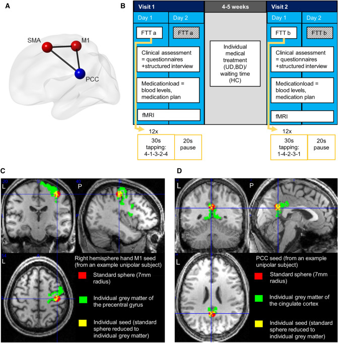
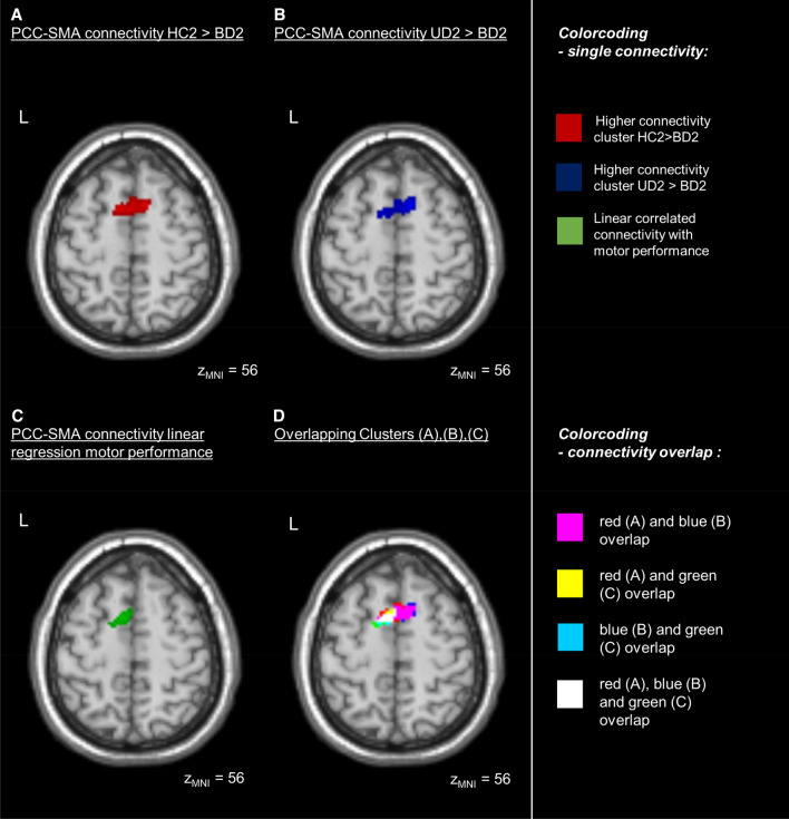
Although implicated in unsuccessful treatment, psychomotor deficits and their neurobiological underpinnings in bipolar (BD) and unipolar (UD) depression remain poorly investigated. Here, we hypothesized that motor performance deficits in depressed patients would relate to basal functional coupling of the hand primary motor cortex (M1) and the posterior cingulate cortex (PCC) with the supplementary motor area (SMA). We performed a longitudinal, naturalistic study in BD, UD and matched healthy controls comprising of two resting-state functional MRI measurements five weeks apart and accompanying assessments of motor performance using a finger tapping task (FTT). A subject-specific seed-based analysis describing functional connectivity between PCC-SMA as well as M1-SMA was conducted. The basal relationships with motor performance were investigated using linear regression models and all measures were compared across groups. Performance in FTT was impaired in BD in comparison to HC in both sessions. Behavioral performance across groups correlated significantly with resting state functional coupling of PCC-SMA, but not of M1-SMA regions. This relationship was partially reflected in a reduced PCC-SMA connectivity in BD vs HC in the second session. Exploratory evaluation of large-scale networks coupling (SMN-DMN) exhibited no correlation to motor performance. Our results shed new light on the association between the degree of disruption in the SMA-PCC anticorrelation and the level of motor impairment in BD.
Keywords: Bipolar disorder; Finger tapping; Major depressive disorder; Posterior cingulate cortex; Resting state functional magnetic resonance imaging; Supplementary motor area.
© 2023. The Author(s).
Conflict of interest statement
The authors report no competing interests.
Figures


Similar articles
-
Altered effective connectivity model in the default mode network between bipolar and unipolar depression based on resting-state fMRI.J Affect Disord. 2015 Aug 15;182:8-17. doi: 10.1016/j.jad.2015.04.009. Epub 2015 Apr 11. J Affect Disord. 2015. PMID: 25942576
-
Interhemispheric resting state functional connectivity abnormalities in unipolar depression and bipolar depression.Bipolar Disord. 2015 Aug;17(5):486-95. doi: 10.1111/bdi.12315. Epub 2015 Jun 9. Bipolar Disord. 2015. PMID: 26241359
-
Functional connectivity of supplementary motor area during finger-tapping in major depression.Compr Psychiatry. 2020 May;99:152166. doi: 10.1016/j.comppsych.2020.152166. Epub 2020 Jan 28. Compr Psychiatry. 2020. PMID: 32182454
-
Differentiating between bipolar and unipolar depression in functional and structural MRI studies.Prog Neuropsychopharmacol Biol Psychiatry. 2019 Apr 20;91:20-27. doi: 10.1016/j.pnpbp.2018.03.022. Epub 2018 Mar 28. Prog Neuropsychopharmacol Biol Psychiatry. 2019. PMID: 29601896 Review.
-
Resting-state functional connectivity in individuals with bipolar disorder during clinical remission: a systematic review.J Psychiatry Neurosci. 2018 Aug;43(5):298-316. doi: 10.1503/jpn.170175. J Psychiatry Neurosci. 2018. PMID: 30125243 Free PMC article.
Cited by
-
From pathogenesis of stress-related mental disorders to treatment: beyond the brain.Eur Arch Psychiatry Clin Neurosci. 2024 Apr;274(3):473-474. doi: 10.1007/s00406-024-01791-2. Eur Arch Psychiatry Clin Neurosci. 2024. PMID: 38519738 No abstract available.
-
The unique role of anosognosia in the clinical progression of Alzheimer's disease: a disorder-network perspective.Commun Biol. 2024 Oct 24;7(1):1384. doi: 10.1038/s42003-024-07076-7. Commun Biol. 2024. PMID: 39448784 Free PMC article. Review.
-
Adaptive Whole-Brain Dynamics Predictive Method: Relevancy to Mental Disorders.Research (Wash D C). 2025 Apr 5;8:0648. doi: 10.34133/research.0648. eCollection 2025. Research (Wash D C). 2025. PMID: 40190349 Free PMC article.
-
Characteristic Changes of Prefrontal and Motor Areas in Patients with Type 2 Diabetes and Major Depressive Disorder During a Motor Task of Tai Chi Chuan: A Functional Near-Infrared Spectroscopy Study.Brain Behav. 2024 Oct;14(10):e70071. doi: 10.1002/brb3.70071. Brain Behav. 2024. PMID: 39378277 Free PMC article.
References
MeSH terms
Grants and funding
LinkOut - more resources
Full Text Sources
Medical
