Association of epicardial and intramyocardial fat with ventricular arrhythmias
- PMID: 37640127
- PMCID: PMC10881203
- DOI: 10.1016/j.hrthm.2023.08.033
Association of epicardial and intramyocardial fat with ventricular arrhythmias
Abstract
Background: Among patients with ischemic cardiomyopathy (ICM) and nonischemic cardiomyopathy (NICM), myocardial fibrosis is associated with an increased risk for ventricular arrhythmia (VA). Growing evidence suggests that myocardial fat contributes to ventricular arrhythmogenesis. However, little is known about the volume and distribution of epicardial adipose tissue and intramyocardial fat and their relationship with VAs.
Objective: The purpose of this study was to assess the association of contrast-enhanced computed tomography (CE-CT)-derived left ventricular (LV) tissue heterogeneity, epicardial adipose tissue volume, and intramyocardial fat volume with the risk of VA in ICM and NICM patients.
Methods: Patients enrolled in the PROSE-ICD registry who underwent CE-CT were included. Intramyocardial fat volume (voxels between -180 and -5 Hounsfield units [HU]), epicardial adipose tissue volume (between -200 and -50 HU), and LV tissue heterogeneity were calculated. The primary endpoint was appropriate ICD shocks or sudden arrhythmic death.
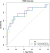
Results: Among 98 patients (47 ICM, 51 NICM), LV tissue heterogeneity was associated with VA (odds ratio [OR] 1.10; P = .01), particularly in the ICM cohort. In the NICM subgroup, epicardial adipose tissue and intramyocardial fat volume were associated with VA (OR 1.11, P = .01; and OR = 1.21, P = .01, respectively) but not in the ICM patients (OR 0.92, P =.22; and OR = 0.96, P =.19, respectively).
Conclusion: In ICM patients, increased fat distribution heterogeneity is associated with VA. In NICM patients, an increased volume of intramyocardial fat and epicardial adipose tissue is associated with a higher risk for VA. Our findings suggest that fat's contribution to VAs depends on the underlying substrate.
Keywords: Contrast-enhanced computed tomography; Epicardial fat; Intramyocardial fat; Ischemic cardiomyopathy; Left ventricular tissue heterogeneity; Nonischemic cardiomyopathy; Ventricular arrhythmia.
Copyright © 2023 Heart Rhythm Society. Published by Elsevier Inc. All rights reserved.
Figures


Comment in
-
Cross talk: Fat and the arrhythmogenic substrate.Heart Rhythm. 2023 Dec;20(12):1706-1707. doi: 10.1016/j.hrthm.2023.09.006. Epub 2023 Sep 12. Heart Rhythm. 2023. PMID: 37709107 No abstract available.
References
-
- Tsao CW, Aday AW, Almarzooq ZI, et al. Heart Disease and Stroke Statistics-2022 Update: A Report From the American Heart Association [published correction appears in Circulation. 2022 Sep 6;146(10):e141]. Circulation. 2022;145(8):e153–e639. - PubMed
-
- Bunch TJ, White RD. Trends in treated ventricular fibrillation in out-of-hospital cardiac arrest: ischemic compared to non-ischemic heart disease. Resuscitation. 2005;67(1):51–54. - PubMed
Publication types
MeSH terms
Grants and funding
LinkOut - more resources
Full Text Sources
Medical

