. 2023 Aug 29;14(1):5250.
doi: 10.1038/s41467-023-40805-w.

Tricolor visible wavelength-selective photodegradable hydrogel biomaterials

Affiliations

Tricolor visible wavelength-selective photodegradable hydrogel biomaterials

Teresa L Rapp et al. Nat Commun. .

Abstract

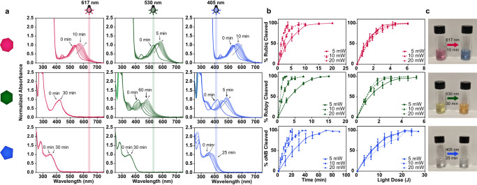
Photodynamic hydrogel biomaterials have demonstrated great potential for user-triggered therapeutic release, patterned organoid development, and four-dimensional control over advanced cell fates in vitro. Current photosensitive materials are constrained by their reliance on high-energy ultraviolet light (<400 nm) that offers poor tissue penetrance and limits access to the broader visible spectrum. Here, we report a family of three photolabile material crosslinkers that respond rapidly and with unique tricolor wavelength-selectivity to low-energy visible light (400-617 nm). We show that when mixed with multifunctional poly(ethylene glycol) macromolecular precursors, ruthenium polypyridyl- and ortho-nitrobenzyl (oNB)-based crosslinkers yield cytocompatible biomaterials that can undergo spatiotemporally patterned, uniform bulk softening, and multiplexed degradation several centimeters deep through complex tissue. We demonstrate that encapsulated living cells within these photoresponsive gels show high viability and can be successfully recovered from the hydrogels following photodegradation. Moving forward, we anticipate that these advanced material platforms will enable new studies in 3D mechanobiology, controlled drug delivery, and next-generation tissue engineering applications.


Conflict of interest statement

The authors declare no competing interests.

Figures

Fig. 1
Fig. 1. Photodegradable crosslinkers cleave in a visible wavelength-selective manner.
a Novel crosslinkers Rubiq (pink heptagon), Rubpy (green hexagon), and oNB (blue pentagon) undergo photolysis. Upon absorption of a photon, each ruthenium complex undergoes ligand exchange via population of a triplet metal-centered state via intersystem crossing from the low-lying metal-to-ligand charge transfer (MLCT) band, resulting in heterolytic ligand/solvent exchange. Crosslinkers are modified with two azides for direct incorporation into hydrogel networks. b Absorbance spectra of Rubiq, Rubpy, and oNB. Each crosslinker exhibits an extended absorbance spectrum beyond their λmax permitting excitation at wavelengths lower than λmax. c PEG-based hydrogel formation occurs spontaneously upon mixing each crosslinker with 4-arm PEG-BCN via strain-promoted azide-alkyne cycloaddition. Resulting hydrogels degrade under red, green, or blue light with perfect unidirectional orthogonality (>617 nm for Rubiq, >530 nm for Rubpy, and >405 nm for oNB).
Fig. 2
Fig. 2. Photolabile crosslinkers can be selectively cleaved with differently colored visible light.
a Rubiq (pink heptagon), Rubpy (green hexagon), and oNB (blue pentagon) individually subjected to 617, 530, and 405 nm light (10 mW cm−2, 0–60 min) undergo wavelength-selective photolysis, indicated by absorbance spectral changes. b Photolysis proceeds efficiently with varied power (5, 10, 20 mW) in a light dose-dependent manner. Here, light dosage is calculated as the product of light intensity and exposure time. n = 3 for each photolysis, error bars represent standard deviation about the experimental mean. c Color change was observed for Rubiq and Rubpy upon irradiation (10 mW, varied wavelengths and exposure times) due to the significant absorbance shift following ligand exchange.
Fig. 3
Fig. 3. Wavelength-orthogonal hydrogel degradation via red, green, and blue light.
a Hydrogels formed via SPAAC chemistry between PEG-tetraBCN and the azide-flanked crosslinkers. b Rubiq (pink heptagon)-, Rubpy (green hexagon)-, and oNB (blue pentagon)-crosslinked hydrogels form equivalently stiff hydrogels (G’ ~ 2-3 kPa) as determined rheometrically. c Individually crosslinked hydrogels are unidirectionally orthogonally degraded, with stable storage modulus observed in Rubpy-crosslinked gels under red light irradiation and oNB-crosslinked gels under red and green light irradiation. d Hydrogels were formed with equal ratios of all three photolabile crosslinkers and increasing amounts of nondegradable diazido-tri(ethylene glycol) (TEG) crosslinker (0–90%, white rectangle). e Intermediate stiffnesses were accessible through partial degradation of hydrogel crosslinks. Hydrogels were exposed to red, green, then blue light (35 min each, 10 mW cm2) to selectively cleave specific amounts of crosslinks within the material. Reported percentages refer to the amount of stable, non-cleavable crosslink present in the material. f Spatial control over hydrogel degradation demonstrated by the casting of individually crosslinked hydrogels in close proximity. Open microfluidic methodologies were used to cast interconnected multimaterial geometries; exposure to red light resulted in the center Rubiq degradation, observed after a 60- min wash. Subsequent exposures to green and blue light rapidly degraded the Rubpy and oNB portions, respectively. Scale bar = 5 mm. g Rubiq hydrogels were completely degraded through up to 2 cm of complex tissue (skin-on pork belly shown here), Rubpy hydrogels through 1 cm, and oNB (dyed with Cy5) through 0.5 cm.
Fig. 4
Fig. 4. Cell viability following encapsulation in and photorelease from Rubiq, Rubpy, and oNB-crosslinked hydrogels.
a Live/dead staining of 10T1/2 fibroblasts encapsulated in individually crosslinked hydrogels, shown as single z-slice obtained via confocal micrography. Calcein (green) and ethidium homodimer (EtHD, red) dyes were used for live and dead staining respectively. Scale bar = 50 μm. b Encapsulated cell viability at 24 h post encapsulation (cell counts collected from 40 μm x 1 mm × 1 mm section through the vertical center of the hydrogel). Cell counting revealed no statistical difference (p > 0.05, one-way ANOVA) in cell viability across all crosslinkers at 24 h. n = 3 individual biological replicates, error bars represent standard deviation about the experimental mean. c Rubiq, Rubpy, and oNB hydrogels were exposed to red, green, or blue light respectively (617 nm, 530 nm, 405 nm; 10 mW cm2) and the released fibroblasts allowed to settle to the bottom of the well over 24 h prior to imaging. Surviving cells exhibited classical fibroblast morphology upon calcein green/EtHD staining. Scale bars = 50 μm. d High levels of viable cells were recovered from each hydrogel formulation, though cells released from Rubiq hydrogels showed lower levels of viability compared to Rubpy and oNB. **p = 0.0036 by one-way ANOVA, n = 3 individual biological replicates, error bars represent standard deviation about the experimental mean. e mCherry+, GFP+, and BFP+ hS5 cells were encapsulated in Rubiq, Rubpy, and oNB hydrogels respectively. 24 h after encapsulation, hydrogels were exposed to 617 nm, 530 nm, or 405 nm light. f Images of hydrogels bearing mCherry+, GFP+, or BFP+ hS5 cells following light exposure. Scale bars = 500 μm. Experiment performed in biological triplicate with similar results; representative images given. g Flow cytometry histograms show only expected cell populations were released into the media, demonstrating the orthogonality of the hydrogel system.

References

    1. Ruskowitz ER, DeForest CA. Photoresponsive biomaterials for targeted drug delivery and 4D cell culture. Nat. Rev. Mater. 2018;3:17087.
    1. Rapp TL, DeForest CA. Visible Light‐Responsive Dynamic Biomaterials: Going Deeper and Triggering More. Adv. Healthc. Mater. 2020;9:1901553. - PubMed
    1. Rapp, T. L. & DeForest, C. A. Targeting drug delivery with light: A highly focused approach. Adv. Drug Deliv. Rev. 171, 94–107 (2021). - PMC - PubMed
    1. Munoz-Robles BG, Kopyeva I, DeForest CA. Biomaterials: Surface Patterning of Hydrogel Biomaterials to Probe and Direct Cell–Matrix Interactions (Adv. Mater. Interfaces 21/2020) Adv. Mater. Interf. 2020;7:2070116.
    1. Gilbert PM, et al. Substrate elasticity regulates skeletal muscle stem cell self-renewal in culture. Science. 2010;329:1078–1081. - PMC - PubMed

Publication types