. 2024 Jan;45(1):137-149.
doi: 10.1038/s41401-023-01148-w. Epub 2023 Aug 28.

Sirtuin 6 protects against podocyte injury by blocking the renin-angiotensin system by inhibiting the Wnt1/β-catenin pathway

Affiliations

Sirtuin 6 protects against podocyte injury by blocking the renin-angiotensin system by inhibiting the Wnt1/β-catenin pathway

Hua Miao et al. Acta Pharmacol Sin. 2024 Jan.

Abstract

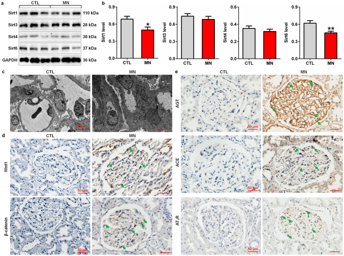
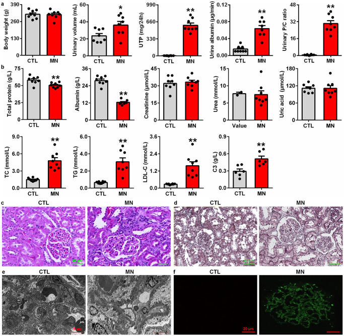
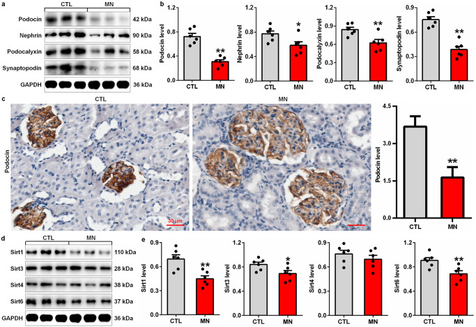
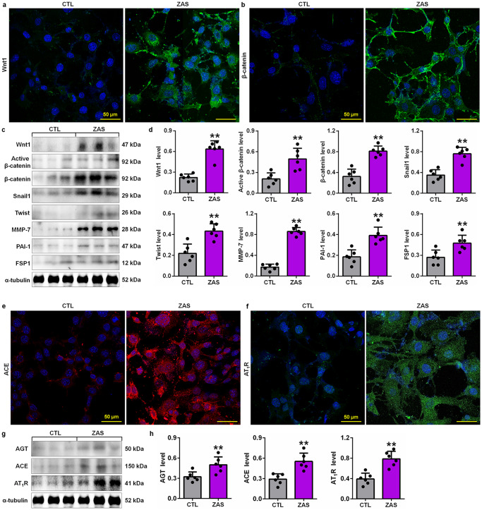
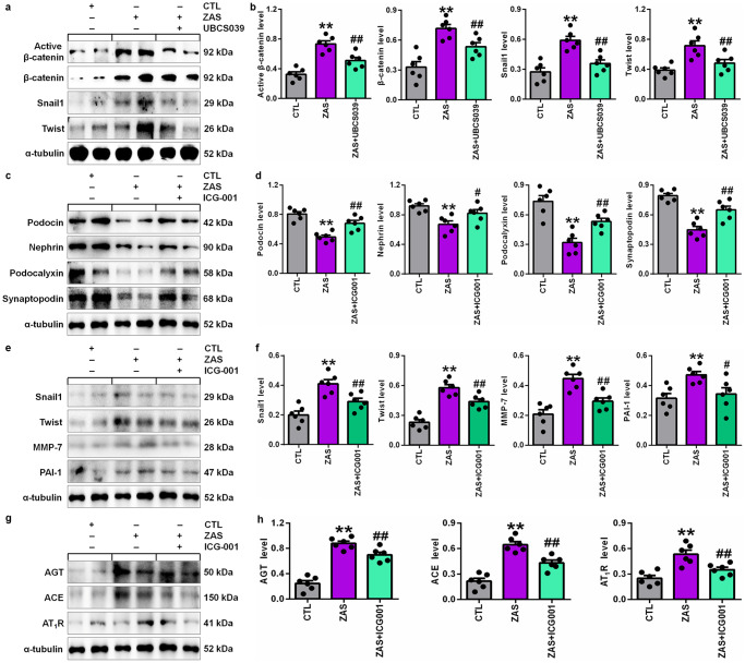
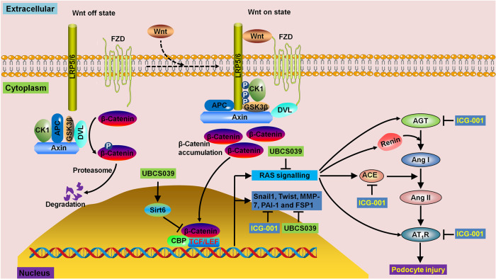
Sirtuins (Sirts) are a family of nicotinamide adenine dinucleotide-dependent protein deacetylases that share diverse cellular functions. Increasing evidence shows that Sirts play a critical role in podocyte injury, which is a major determinant of proteinuria-associated renal disease. Membranous nephropathy (MN) is a typical glomerular disease in which podocyte damage mediates proteinuria development. In this study we investigated the molecular mechanisms underlying the regulatory roles of Sirt in podocyte injury in MN patients, rats with cationic bovine serum albumin (CBSA)-induced MN and zymosan activation serum (ZAS)-stimulated podocytes. Compared with healthy controls, MN patients showed significant reduction in intrarenal Sirt1 and Sirt6 protein expression. In CBSA-induced MN rats, significant reduction in intrarenal Sirt1, Sirt3 and Sirt6 protein expression was observed. However, only significant decrease in Sirt6 protein expression was found in ZAS-stimulated podocytes. MN patients showed significantly upregulated protein expression of Wnt1 and β-catenin and renin-angiotensin system (RAS) components in glomeruli. CBSA-induced MN rats exhibited significantly upregulated protein expression of intrarenal Wnt1 and β-catenin and their downstream gene products as well as RAS components. Similar results were observed in ZAS-stimulated podocytes. In ZAS-stimulated podocytes, treatment with a specific Sirt6 activator UBCS039 preserved the protein expression of podocin, nephrin and podocalyxin, accompanied by significant inhibition of the protein expression of β-catenin and its downstream gene products, including Snail1 and Twist; treatment with a β-catenin inhibitor ICG-001 significantly preserved the expression of podocyte-specific proteins and inhibited the upregulation of downstream β-catenin gene products accompanied by significant suppression of the protein expression of RAS components. Thus, we demonstrate that Sirt6 ameliorates podocyte injury by blocking RAS signalling via the Wnt1/β-catenin pathway. Sirt6 is a specific therapeutic target for the treatment of podocyte damage-associated renal disease.

Keywords: Sirtuins; UBCS039; Wnt1/β-catenin pathway; membranous nephropathy; podocyte injury; renin-angiotensin system.


Conflict of interest statement

The authors declare no competing interests.

Figures

Fig. 1
Fig. 1. Intrarenal Sirt deficiency, a hyperactive Wnt1/β-catenin pathway and RAS overexpression in MN patients.
a Protein expression levels of intrarenal Sirt1, Sirt3, Sirt4 and Sirt6 in controls and patients with MN. b Quantitative analysis of the protein expression levels of intrarenal Sirt1, Sirt3, Sirt4 and Sirt6 in controls and patients with MN. *P < 0.05, **P < 0.01 compared with CTL. c Electron microscopy images of kidney tissues from controls and patients with MN. d Immunohistochemical analysis of intrarenal Wnt1 and β-catenin expression in controls and patients with MN. e Immunohistochemical analysis of intrarenal AGT, ACE and AT1R expression in controls and patients with MN. Arrows indicate protein expression.
Fig. 2
Fig. 2. Renal function decline and renal injury in CBSA-induced rats.
a Body weight, urine volume, UTP, urine albumin excretion rate and the urinary protein/creatinine ratio in control and MN rats. b Levels of total protein, albumin, creatinine, urea, uric acid, TC, TG, LDL-C and C3 in control and MN rats. c Images of H&E staining of kidney tissues in control and MN rats. d Images of PASM staining of the kidney tissues in control and MN rats. e Electron microscopy images of the kidney tissues of control and MN rats. f Images of IgG4 immunofluorescence staining of the kidney tissues of control and MN rats. *P < 0.05, **P < 0.01 compared with CTL rats.
Fig. 3
Fig. 3. Podocyte injury and downregulated protein expression of Sirts in CBSA-induced rats.
a Protein expression levels of intrarenal podocin, nephrin, podocalyxin and synaptopodin in control and CBSA-induced MN rats. b Quantitative analysis of the protein expression of intrarenal podocin, nephrin, podocalyxin and synaptopodin in control and CBSA-induced MN rats. c Immunohistochemical analysis of intrarenal podocin expression in control and CBSA-induced MN rats. d Protein expression levels of intrarenal Sirt1, Sirt3, Sirt4 and Sirt6 in control and CBSA-induced MN rats. e Quantitative analysis of the protein expression levels of intrarenal Sirt1, Sirt3, Sirt4 and Sirt6 in control and CBSA-induced MN rats. *P < 0.05, **P < 0.01 compared with CTL rats.
Fig. 4
Fig. 4. Activation of the Wnt1/β-catenin pathway and RAS signalling in CBSA-induced rats.
a Immunohistochemical analysis of intrarenal Wnt1 expression in control and CBSA-induced MN rats. b Immunohistochemical analysis of intrarenal β-catenin expression in control and CBSA-induced MN rats. c Protein expression levels of intrarenal Wnt1, active β-catenin and β-catenin and the downstream gene products, including Snail1, Twist, MMP-7, PAI-1 and FSP1, in control and CBSA-induced MN rats. d Quantitative analysis of the protein expression of intrarenal Wnt1, active β-catenin, β-catenin Snail1, Twist, MMP-7, PAI-1 and FSP1 in control and CBSA-induced MN rats. e Immunohistochemical analysis of intrarenal ACE expression in control and CBSA-induced MN rats. f Immunohistochemical analysis of intrarenal AT1R expression in control and CBSA-induced MN rats. g Protein expression levels of intrarenal AGT, ACE and AT1R in control and CBSA-induced MN rats. h Quantitative analysis of the protein expression of intrarenal AGT, ACE and AT1R in control and CBSA-induced MN rats. *P < 0.05, **P < 0.01 compared with CTL rats.
Fig. 5
Fig. 5. Sirt6 deficiency exacerbated podocyte injury in ZAS-stimulated podocytes.
a Protein expression levels of podocin, nephrin, podocalyxin and synaptopodin in control and ZAS-stimulated podocytes. b Quantitative analysis of the protein expression of podocin, nephrin, podocalyxin and synaptopodin in control and ZAS-stimulated podocytes. c Protein expression levels of intrarenal Sirt1, Sirt3, Sirt4 and Sirt6 in control and ZAS-stimulated podocytes. d Quantitative analysis of the protein expression levels of intrarenal Sirt1, Sirt3, Sirt4 and Sirt6 in control and ZAS-stimulated podocytes. e Protein expression levels of Sirt6, podocin, nephrin and podocalyxin in ZAS-stimulated podocytes treated with UBCS039. f Quantitative analysis of the protein expression levels of Sirt6, podocin, nephrin and podocalyxin in ZAS-stimulated podocytes treated with UBCS039. *P < 0.05, **P < 0.01 compared with CTL; ##P < 0.01 compared with ZAS-stimulated podocytes.
Fig. 6
Fig. 6. Activation of the Wnt1/β-catenin pathway and RAS signalling in ZAS-stimulated podocytes.
a Images of Wnt1 immunofluorescence staining in control and ZAS-stimulated podocytes. b Images of β-catenin immunofluorescence staining in control and ZAS-stimulated podocytes. c Protein expression levels of Wnt1, active β-catenin and β-catenin and its downstream gene products, including Snail1, Twist, MMP-7, PAI-1 and FSP1, in control and ZAS-stimulated podocytes. d Quantitative analysis of the protein expression of Wnt1, active β-catenin, β-catenin Snail1, Twist, MMP-7, PAI-1 and FSP1 in control and ZAS-stimulated podocytes. e Images of ACE immunofluorescence staining in control and ZAS-stimulated podocytes. f Images of AT1R immunofluorescence staining in control and ZAS-stimulated podocytes. g Protein expression levels of AGT, ACE and AT1R in control and ZAS-stimulated podocytes. h Quantitative analysis of the protein expression of AGT, ACE and AT1R in control and ZAS-stimulated podocytes. **P < 0.01 compared with CTL rats.
Fig. 7
Fig. 7. Sirt6 reduced podocyte injury by blocking RAS signalling via the Wnt1/β-catenin pathway.
a Protein expression levels of active β-catenin, β-catenin, Snail1 and Twist in ZAS-stimulated podocytes treated with UBCS039. b Quantitative analysis of the protein expression levels of active β-catenin, β-catenin, Snail1 and Twist in ZAS-stimulated podocytes treated with UBCS039. c Protein expression levels of podocin, nephrin, podocalyxin and synaptopodin in ZAS-stimulated podocytes treated with ICG-001. d Quantitative analysis of the protein expression of podocin, nephrin, podocalyxin and synaptopodin in ZAS-stimulated podocytes treated with ICG-001. e Protein expression levels of Snail1, Twist, MMP-7 and PAI-1 in ZAS-stimulated podocytes treated with ICG-001. f Quantitative analysis of the protein expression of Snail1, Twist, MMP-7 and PAI-1 in ZAS-stimulated podocytes treated with ICG-001. g Protein expression levels of AGT, ACE and AT1R in ZAS-stimulated podocytes treated with ICG-001. h Quantitative analysis of the protein expression of AGT, ACE and AT1R in ZAS-stimulated podocytes treated with ICG-001. **P < 0.01 compared with CTL; #P < 0.05, ##P < 0.01 compared with ZAS-stimulated podocytes.
Fig. 8
Fig. 8. Summary of the underlying molecular mechanism of Sirt6 deficiency, the hyperactive Wnt1/β-catenin pathway and RAS overexpression in podocyte injury.
Sirt6 deficiency, a hyperactive Wnt1/β-catenin pathway and RAS overexpression are observed in patients with MN, CBSA-injected rats and ZAS-stimulated podocytes. UBCS039 treatment significantly inhibits the protein expression of β-catenin and its downstream gene products, including Snail1 and Twist, in ZAS-stimulated podocytes. Treatment with ICG-001 significantly inhibits the protein expression of downstream β-catenin gene products, including Snail1, Twist, MMP-7 and PAI-1, which is accompanied by significantly downregulated protein expression of AGT, ACE and AT1R in ZAS-stimulated podocytes. Podocyte injury is also ameliorated by treatment with UBCS039 and ICG-001.

References

    1. Morigi M, Perico L, Benigni A. Sirtuins in renal health and disease. J Am Soc Nephrol. 2018;29:1799–809. doi: 10.1681/ASN.2017111218. - DOI - PMC - PubMed
    1. Qi W, Hu C, Zhao D, Li X. SIRT1-SIRT7 in diabetic kidney disease: biological functions and molecular mechanisms. Front Endocrinol (Lausanne) 2022;13:801303. doi: 10.3389/fendo.2022.801303. - DOI - PMC - PubMed
    1. Liu T, Yang L, Mao H, Ma F, Wang Y, Li S, et al. Sirtuins as novel pharmacological targets in podocyte injury and related glomerular diseases. Biomed Pharmacother. 2022;155:113620. doi: 10.1016/j.biopha.2022.113620. - DOI - PubMed
    1. Zhong Y, Lee K, He JC. SIRT1 is a potential drug target for treatment of diabetic kidney disease. Front Endocrinol (Lausanne) 2018;9:624. doi: 10.3389/fendo.2018.00624. - DOI - PMC - PubMed
    1. Li P, Liu Y, Qin X, Chen K, Wang R, Yuan L, et al. SIRT1 attenuates renal fibrosis by repressing HIF-2α. Cell Death Discov. 2021;7:59. doi: 10.1038/s41420-021-00443-x. - DOI - PMC - PubMed