. 2023 Nov;202(2):397-408.
doi: 10.1007/s10549-023-07090-z. Epub 2023 Aug 28.

FOXO1-regulated lncRNA CYP1B1-AS1 suppresses breast cancer cell proliferation by inhibiting neddylation

Affiliations

FOXO1-regulated lncRNA CYP1B1-AS1 suppresses breast cancer cell proliferation by inhibiting neddylation

Li Tang et al. Breast Cancer Res Treat. 2023 Nov.

Abstract

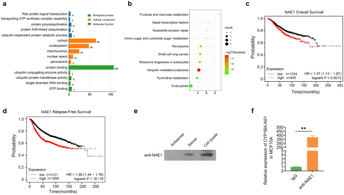
Purpose: Overactivated neddylation is considered to be a common event in cancer. Long non-coding RNAs (lncRNAs) can regulate cancer development by mediating post-translational modifications. However, the role of lncRNA in neddylation modification remains unclear.

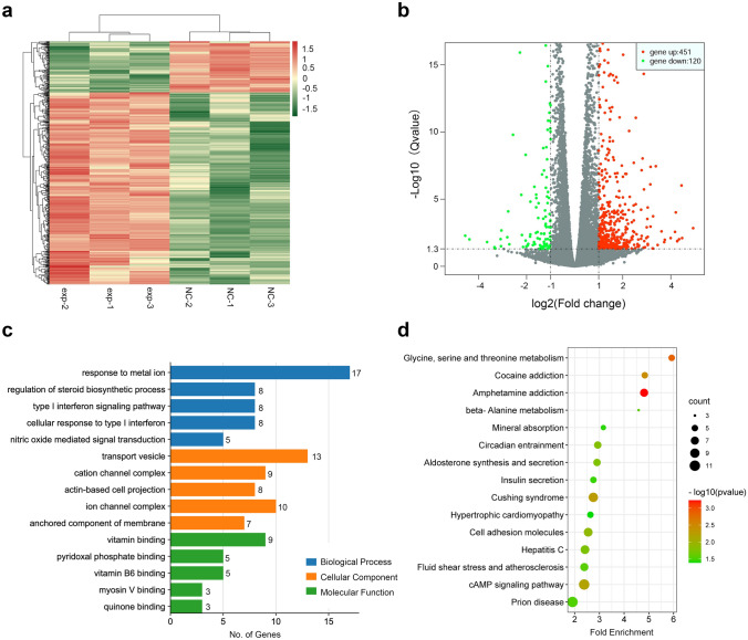
Methods: LncRNA cytochrome P450 family 1 subfamily B member 1 antisense RNA 1 (CYP1B1-AS1) expression in breast cancer tissues was evaluated by RT-PCR and TCGA BRCA data. Gain and loss of function experiments were performed to explore the role of CYP1B1-AS1 in breast cancer cell proliferation and apoptosis in vitro and in vivo. Luciferase assay, CHIP-qPCR assay, transcriptome sequencing, RNA-pulldown assay, mass spectrometry, RIP-PCR and Western blot were used to investigate the regulatory factors of CYP1B1-AS1 expression and the molecular mechanism of CYP1B1-AS1 involved in neddylation modification.

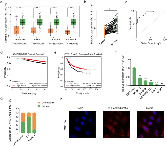
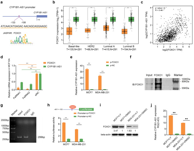
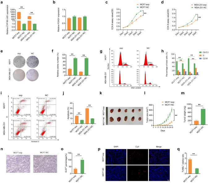
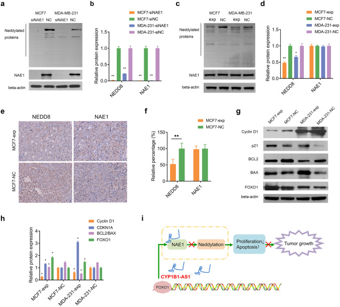
Results: We found that CYP1B1-AS1 was down-regulated in breast cancer tissues and correlated with prognosis. In vivo and in vitro functional experiments confirmed that CYP1B1-AS1 inhibited cell proliferation and induced apoptosis. Mechanistically, CYP1B1-AS1 was regulated by the transcription factor, forkhead box O1 (FOXO1), and could be upregulated by inhibiting the PI3K/FOXO1 pathway. Moreover, CYP1B1-AS1 bound directly to NEDD8 activating enzyme E1 subunit 1 (NAE1) to regulate protein neddylation.

Conclusion: This study reports for the first time that CYP1B1-AS1 inhibits protein neddylation to affect breast cancer cell proliferation, which provides a new strategy for the treatment of breast cancer by lncRNA targeting neddylation modification.

Keywords: Breast cancer; FOXO1; Long noncoding RNA; NAE1; NEDD8; Neddylation.


Conflict of interest statement

The authors declare that they have no conflict of interest.

Figures

Fig. 1
Fig. 1
Downregulation of CYP1B1-AS1 is associated with breast cancer progression. a The Cancer Genome Atlas (TCGA) data analysis of CYP1B1-AS1 expression in each molecular subtype of breast cancer. N, non-tumor tissue. T, tumor tissue. **P < 0.01. b Quantitative real-time polymerase chain reaction (qPCR) was used to analyze the expression level of CYP1B1-AS1 in breast cancer tissues and adjacent non-tumor tissues of 44 patients. ***P < 0.001. c Receiver operating characteristic curve analysis of the diagnostic value of CYP1B1-AS1 in 44 paired breast tissue samples. P < 0.001. d Kaplan–Meier Plotter analysis of the relationship between CYP1B1-AS1 expression and patients’ overall survival. Kaplan–Meier Plotter uses data from public databases such as GEO and TCGA and provides the best available cut-off value. The cut-off value used in the analysis is 4. P < 0.001. e Kaplan–Meier Plotter analysis of the relationship between CYP1B1-AS1 expression and patients’ recurrence-free survival. Kaplan–Meier Plotter uses data from public databases such as GEO and TCGA and provides the best available cut-off value. The cut-off value used in the analysis is 1.28. P < 0.001. f qPCR was used to analyze the expression level of CYP1B1-AS1 in breast cancer cells (BT-549, SK-BR-3, MDA-MB-231, T-47D, and MCF7), and the mammary epithelial cell, MCF10A, was used as control. **P < 0.01. g Nuclear/cytoplasmic separation combined with qPCR were used to analyze the distribution of CYP1B1-AS1 in MCF10A cells. U6 and GAPDH were used as the reference for nuclear RNA extraction and cytoplasmic RNA extraction, respectively. h Fluorescence in situ hybridization (FISH) experiments combined with laser confocal microscopy showed the distribution of CYP1B1-AS1 in MCF10A cells (× 1000)
Fig. 2
Fig. 2
The upstream regulatory mechanism of CYP1B1-AS1. a Schematic representation of the binding sites of FOXO1 predicted by JASPAR in the CYP1B1-AS1 promoter region. b The Cancer Genome Atlas (TCGA) data were analyzed to determine the expression of FOXO1 in various molecular subtypes of breast cancer. N, non-tumor tissue. T, tumor tissue. **P < 0.01. c TCGA data were analyzed to determine the correlation between CYP1B1-AS1 and FOXO1 expression in breast cancer. R, Pearson correlation coefficient. P < 0.001. d Quantitative real-time polymerase chain reaction (qPCR) analysis of the effect of interfering FOXO1 on CYP1B1-AS1 expression in MCF10A cells. FsiRNA, FOXO1 siRNA. siNC, negative control for siRNA. **P < 0.01. e qPCR analysis of the effect of up-regulated FOXO1 expression on CYP1B1-AS1 expression in MCF7 and MDA-MB-231 cells. p-FOXO1, pcDNA3.1-FOXO1. p-NC, negative control for pcDNA3.1. **P < 0.01. f Western blot results of chromatin immunoprecipitation (CHIP) assay to detect FOXO1 binding to CYP1B1-AS1 promoter fragment. Input, positive control. IgG, negative control. g Gel electropherogram of the amplified promoter fragment in CHIP-qPCR assay. Input, positive control. IgG, negative control. h Dual-luciferase assay was used to detect the downstream regulation of FOXO1 binding to the promoter fragment. p-FOXO1, pcDNA3.1-FOXO1. p-NC, negative control for pcDNA3.1. **P < 0.01. i Western blot was used to detect the changes in FOXO1 protein expression after LY294002 treatment. LY, LY294002. Solvent DMSO as blank control. j qPCR was used to detect changes in CYP1B1-AS1 expression after LY294002 treatment. LY, LY294002. Solvent DMSO as blank control. **P < 0.01
Fig. 3
Fig. 3
Upregulation of CYP1B1-AS1 inhibits cell proliferation and induces apoptosis. a Quantitative real-time polymerase chain reaction (qPCR) was used to detect the changes of CYP1B1-AS1 after lentivirus infection of MCF7 and MDA-MB-231 cells. **P < 0.01. b qPCR was used to detect FOXO1 mRNA in MCF7 and MDA-MB-231 cells after up-regulation of CYP1B1-AS1. c, d Counting Kit 8 (CCK-8) was used to detect the changes in the proliferation ability of MCF7 and MDA-MB-231 cells after up-regulation of CYP1B1-AS1. **P < 0.01. e, f The clone formation assay was used to examine the effect of up-regulation of CYP1B1-AS1 on the clone formation ability of breast cancer single cells. **P < 0.01. g, h Flow cytometry was used to analyze the effect of up-regulation of CYP1B1-AS1 on breast cancer cell cycle. **P < 0.01. i, j Flow cytometry was used to analyze the effect of up-regulation of CYP1B1-AS1 on apoptosis. **P < 0.01. k, m Representative images of tumor tissue in the xenograft model and compared the volume and weight of tumor tissue in the MCF7-exp and MCF7-NC groups (n = 6). Tumor volumes shown in l were calculated every 3 days after injection. **P < 0.01. n, o Representative images of immunohistochemical staining showing the expression levels of Ki-67 in MCF7-exp and MCF7-NC transplanted tumor tissues (× 400). **P < 0.01. p, q Representative images of TUNEL fluorescence staining showing the level of apoptosis in MCF7-exp and MCF7-NC transplanted tumor tissues (× 400). **P < 0.01
Fig. 4
Fig. 4
Transcriptome sequencing analysis of MCF7 cells following CYP1B1-AS1 upregulation. a Cluster heatmap of differential gene expression analysis between MCF7-exp and MCF7-NC groups. b Volcano plots show the distribution of differentially expressed genes between the MCF7-exp and MCF7-NC groups. Q, the corrected P-value. c Horizontal bar graph showing Gene Ontology enrichment analysis of differentially expressed genes, and the top five items with P < 0.05 are shown for each category. d Dot plot showing Kyoto Encyclopaedia of Genes and Genomes pathway enrichment analysis of differentially expressed genes, and the top 15 items with P < 0.05 are shown
Fig. 5
Fig. 5
CYP1B1-AS1 binds directly to NAE1 protein. a Gene Ontology enrichment analysis of CYP1B1-AS1 pull-down proteins, each class showing the top five items of P < 0.05. b Kyoto Encyclopaedia of Genes and Genomes pathway enrichment analysis of CYP1B1-AS1 pull-down proteins, showing all items with P < 0.05. c Kaplan–Meier Plotter analysis of the relationship between NAE1 expression and overall survival in breast cancer patients. P < 0.01. d Kaplan–Meier Plotter analysis of the relationship between NAE1 expression and recurrence-free survival in breast cancer patients. P < 0.001. e Western blot was used to detect NAE1 protein in CYP1B1-AS1 sense strand pull-down protein solution. CYP1B1-AS1 antisense strand pull-down protein solution was used as a negative control, and MCF7-exp whole cell lysate was used as a positive control. f The RNA immunoprecipitation products were isolated and purified, and the amount of CYP1B1-AS1 bound to anti-NAE1 or IgG was measured by qPCR. IgG was used as a negative control. **P < 0.01
Fig. 6
Fig. 6
CYP1B1-AS1 affects neddylation through NAE1. a, b Western blotting was used to detect neddylated proteins in MCF7 and MDA-MB-231 cells after interference with NAE1. siNAE1, NAE1 siRNA. siNC, negative control for siRNA. **P < 0.01. c, d Western blotting was used to detect NAE1 and neddylated proteins in MCF7 and MDA-MB-231 cells after CYP1B1-AS1 upregulation. *P < 0.05, **P < 0.01. e, f Representative images of immunohistochemical staining showing the expression of NEDD8 and NAE1 in xenograft tumors. **P < 0.01. g, h Western blot was used to detect the changes in cyclin D1, p21, BCL2, BAX and FOXO1 after up-regulation of CYP1B1-AS1. *P < 0.05. i Schematic diagram of lncRNA CYP1B1-AS1 inhibiting the proliferation of breast cancer by binding NAE1

Similar articles

Cited by

References

    1. Britt KL, Cuzick J, Phillips KA. Key steps for effective breast cancer prevention. Nat Rev Cancer. 2020;20:417–436. doi: 10.1038/s41568-020-0266-x. - DOI - PubMed
    1. Jin H, Du W, Huang W, Yan J, Tang Q, Chen Y, Zou Z. lncRNA and breast cancer: progress from identifying mechanisms to challenges and opportunities of clinical treatment. Mol Ther Nucl acids. 2021;25:613–637. doi: 10.1016/j.omtn.2021.08.005. - DOI - PMC - PubMed
    1. Verhoeff TJ, Holloway AF, Dickinson JL. A novel long non-coding RNA regulates the integrin, ITGA2 in breast cancer. Breast Cancer Res Treat. 2022;192:89–100. doi: 10.1007/s10549-021-06496-x. - DOI - PubMed
    1. Zhou L, Jiang Y, Luo Q, Li L, Jia L. Neddylation: a novel modulator of the tumor microenvironment. Mol Cancer. 2019;18:77. doi: 10.1186/s12943-019-0979-1. - DOI - PMC - PubMed
    1. Watson IR, Irwin MS, Ohh M. NEDD8 pathways in cancer, Sine Quibus Non. Cancer Cell. 2011;19:168–176. doi: 10.1016/j.ccr.2011.01.002. - DOI - PubMed