. 2023 Dec;36(6):2635-2647.
doi: 10.1007/s10278-023-00871-4. Epub 2023 Aug 28.

Enhancing Caries Detection in Bitewing Radiographs Using YOLOv7

Affiliations

Enhancing Caries Detection in Bitewing Radiographs Using YOLOv7

Wannakamon Panyarak et al. J Digit Imaging. 2023 Dec.

Abstract

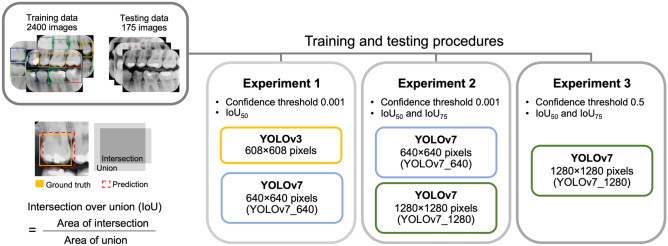
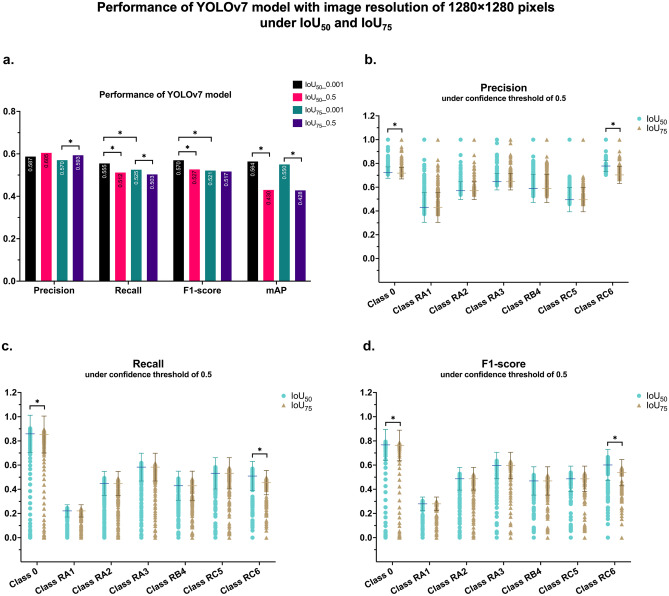
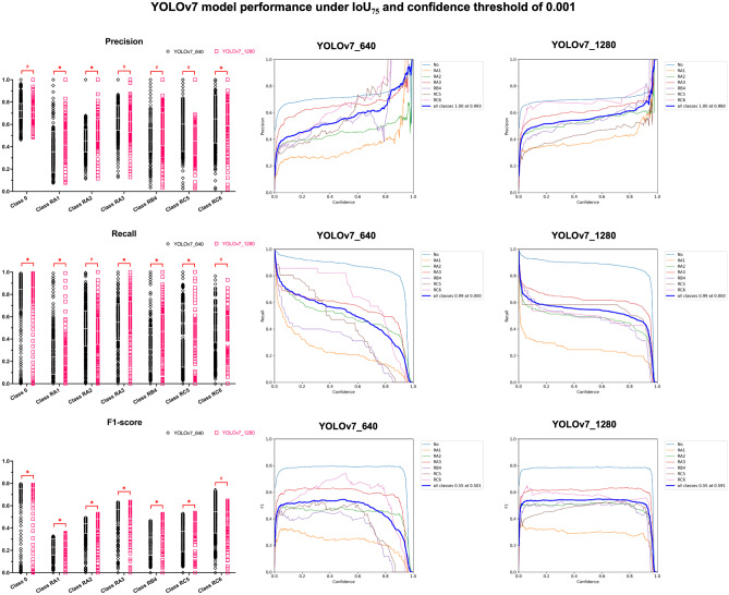
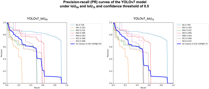
The study aimed to evaluate the impact of image size, area of detection (IoU) thresholds and confidence thresholds on the performance of the YOLO models in the detection of dental caries in bitewing radiographs. A total of 2575 bitewing radiographs were annotated with seven classes according to the ICCMS radiographic scoring system. YOLOv3 and YOLOv7 models were employed with different configurations, and their performances were evaluated based on precision, recall, F1-score and mean average precision (mAP). Results showed that YOLOv7 with 640 × 640 pixel images exhibited significantly superior performance compared to YOLOv3 in terms of precision (0.557 vs. 0.268), F1-score (0.555 vs. 0.375) and mAP (0.562 vs. 0.458), while the recall was significantly lower (0.552 vs. 0.697). The following experiment found that the overall mAPs did not significantly differ between 640 × 640 pixel and 1280 × 1280 pixel images, for YOLOv7 with an IoU of 50% and a confidence threshold of 0.001 (p = 0.866). The last experiment revealed that the precision significantly increased from 0.570 to 0.593 for YOLOv7 with an IoU of 75% and a confidence threshold of 0.5, but the mean-recall significantly decreased and led to lower mAPs in both IoUs. In conclusion, YOLOv7 outperformed YOLOv3 in caries detection and increasing the image size did not enhance the model's performance. Elevating the IoU from 50% to 75% and confidence threshold from 0.001 to 0.5 led to a reduction of the model's performance, while simultaneously improving precision and reducing recall (minimizing false positives and negatives) for carious lesion detection in bitewing radiographs.

Keywords: Bitewing radiograph; Caries detection; Confidence threshold; Dental caries; Detection area.


Conflict of interest statement

The authors declare no competing interests.

Figures

Fig. 1
Fig. 1
Schematic diagram illustrates intersection over union (IoU) operations and the experiments conducted in this study
Fig. 2
Fig. 2
Depicts the validation and testing outcomes comparing YOLOv3 for 608 × 608 pixels and YOLOv7 for 640 × 640 pixels (YOLOv7_640) input bitewing radiographs at an IoU of 50 and a confidence threshold of 0.001. The asterisk (*) denotes a statistically significant increase in precision, F1-score and mean average precision (mAP) achieved by YOLOv7_640. Conversely, the hash symbol (#) denotes a statistically significant decrease in recall for YOLOv7_640
Fig. 3
Fig. 3
Displays the mean average precision (mAP) of YOLOv7 models with an Intersection over Union (IoU) of 50 and 75, at a confidence threshold of 0.001. The mAP is measured for bitewing radiographs with input resolutions of 640 × 640 (YOLOv7_640) and 1280 × 1280 pixels (YOLOv7_1280)
Fig. 4
Fig. 4
Depicts bar graphs illustrating the precision, recall, F1-score and mAP of the YOLOv7 model using an image input size of 1280 × 1280 pixels. The evaluations are performed under IoU50 and IoU75 with confidence thresholds of 0.001 and 0.5 (a). Additionally, the precision, recall and F1-score plots (b, c, d) representing the mAP for caries detection in each ICCMS classification are presented for both IoUs. The results reveal significant differences in class 0 and class RC6, denoted by the asterisk (*) notation
Fig. 5
Fig. 5
Displays examples of bitewing radiographs with bounding boxes that were annotated by radiologists based on the ICCMS radiographic scoring system (a, d, g). The middle column (b, e, h) demonstrates the bitewing radiographs with bounding boxes that were created by the YOLOv7 model with IoU50 and a confidence threshold of 0.001, This resulted in multiple bounding boxes covering each tooth, indicating that the model kept all the predictions with a confidence score above 0.1%. The right column (c, f, i) shows the bitewing radiographs with bounding boxes that were created by the model with a confidence threshold of 0.5, resulting in a single bounding box covering each tooth with a confidence score above 50%. It is noteworthy that the model removed the bounding box that covered the left mandibular second molar (i) as the confidence score did not exceed 50%
Fig. 6
Fig. 6
Demonstrates the utilization of Grad-CAM to represent the focal points of the YOLO model. For instance, accurate positive detections are depicted as highlighted red spots in the carious teeth class RC6, specifically in the left mandibular second molar (a) and left maxillary second premolar (b). Conversely, false positive results are observed in the form of a double bounding box for class 0 and RC5, located in the left mandibular canine, where Grad-CAM does not display any highlighted spot (c). Additionally, a false negative result is illustrated in the left mandibular second molar, corresponding to an intense highlighted spot in Grad-CAM (d). However, this radiograph exhibits a false positive result in the left maxillary second molar as class RA2, attributed to the presence of restoration
Fig. 7
Fig. 7
Presents scattered plots illustrating the precision, recall and F1-score of the YOLOv7 models under the IoU50 and confidence threshold of 0.001. Additionally, the performance plots for both YOLOv7_640 and YOLOv7_1280, corresponding to input bitewing radiographs of 640 × 640 and 1280 × 1280 pixels, respectively, are shown. These evaluations are specifically conducted for caries detection, utilizing the ICCMS radiographic scoring system. In the plots, significantly higher values of YOLOv7_1280 are denoted by an asterisk (*), while significantly lower values are indicated by the hash symbol (#). The confidence thresholds range from 0 to 1.0, allowing for a comprehensive analysis of the model's performance
Fig. 8
Fig. 8
Illustrates scatter plots showcasing the precision, recall and F1-score of YOLOv7 models, considering an IoU threshold of 75% (IoU75) and a confidence threshold of 0.001. Additionally, the performance plots, encompassing confidence thresholds ranging from 0 to 1.0, are presented for both input bitewing radiographs of 640 × 640 (YOLOv7_640) and 1280 × 1280 (YOLOv7_1280) pixels, specifically for caries detection using the ICCMS radiographic scoring system. In the figure, an asterisk (*) denotes significantly higher values observed for YOLOv7_1280, while a hash symbol (#) indicates significantly lower values observed for YOLOv7_1280
Fig. 9
Fig. 9
Illustrates the precision-recall (PR) curves of the YOLOv7 model under two different Intersection over Union (IoU) thresholds, namely 50% (IoU50) and 75% (IoU75), while utilizing a confidence threshold of 0.5. The analysis reveals significant differences between these two IoUs in the performance of class 0 and class RC6. Specifically, the PR curves associated with IoU75 exhibit significantly lower values, leading to a reduced mean average precision (mAP)

Similar articles

Cited by

References

    1. Righolt AJ, Jevdjevic M, Marcenes W, Listl S. Global-, Regional-, and Country-Level Economic Impacts of Dental Diseases in 2015. J Dent Res. 2018;97(5):501–507. doi: 10.1177/0022034517750572. - DOI - PubMed
    1. Nascimento MM, Bader JD, Qvist V, Litaker MS, Williams OD, Rindal DB, et al. Concordance between preoperative and postoperative assessments of primary caries lesion depth: results from the Dental PBRN. Oper Dent. 2010;35(4):389–396. doi: 10.2341/09-363-C. - DOI - PMC - PubMed
    1. Menem R, Barngkgei I, Beiruti N, Al Haffar I, Joury E. The diagnostic accuracy of a laser fluorescence device and digital radiography in detecting approximal caries lesions in posterior permanent teeth: an in vivo study. Lasers Med Sci. 2017;32:621–628. doi: 10.1007/s10103-017-2157-2. - DOI - PMC - PubMed
    1. Cantu AG, Gehrung S, Krois J, Chaurasia A, Rossi JG, Gaudin R, et al. Detecting caries lesions of different radiographic extension on bitewings using deep learning. J Dent. 2020;100:103425. doi: 10.1016/j.jdent.2020.103425. - DOI - PubMed
    1. Mertens S, Krois J, Cantu AG, Arsiwala LT, Schwendicke F. Artificial intelligence for caries detection: Randomized trial. J Dent. 2021;115:103849. doi: 10.1016/j.jdent.2021.103849. - DOI - PubMed

MeSH terms

LinkOut - more resources