. 2023 Aug 29;9(1):55.
doi: 10.1186/s40981-023-00647-3.

Anesthetic management of scapular Y-osteotomy using a combination of suprascapular nerve block and erector spinae plane block for Sprengel deformity associated with Klippel-Feil syndrome: a case report

Affiliations

Anesthetic management of scapular Y-osteotomy using a combination of suprascapular nerve block and erector spinae plane block for Sprengel deformity associated with Klippel-Feil syndrome: a case report

Mizuho Okada et al. JA Clin Rep. .

Abstract

Background: Klippel-Feil syndrome (KFS) occurs in 1/40,000 individuals and is characterized by cervical fusion. Thirty percent of patients with KFS present with Sprengel deformity, leading to orthopedic problems and limited shoulder abduction. No reports exist regarding anesthetic procedures for pediatric scapular osteotomies.

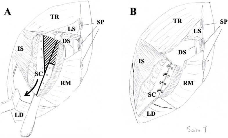
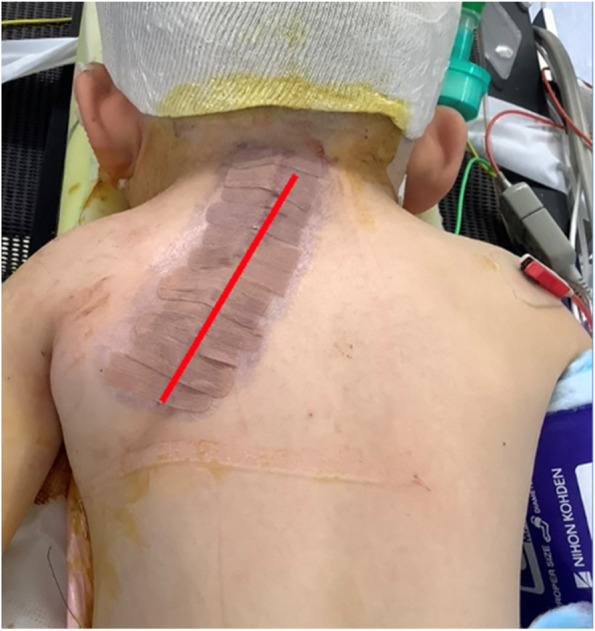
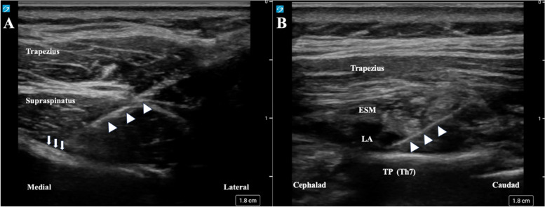
Case presentation: We report a case of a 4-year-and-7-month-old boy (95.6 cm, 14.7 kg) who underwent left scapular osteotomy. At the age of 8 months, he also underwent a right lower lobectomy due to a congenital pulmonary airway malformation. We decided to use a combination of suprascapular nerve block (SSNB), erector spinae plane block (ESPB), and general anesthesia. He received regular acetaminophen administration and fentanyl 5-10 μg/hour intravenously until 20 h postoperatively and remained on ≤ 2/10 in the Wong-Baker Face Scale (0: no hurt; 10: hurts worst).

Conclusion: The combination of SSNB and ESPB could be an option for perioperative analgesia for scapular osteotomies.

Keywords: Erector spinae plane block; Pediatric anesthesia; Peripheral nerve block; Suprascapular nerve block.


Conflict of interest statement

The authors declare that they have no competing interests.

Figures

Fig. 1
Fig. 1
Surgical schematic of scapular Y-osteotomy proposed by Saisu (handwritten by himself). A Solid lines indicate the lines of osteotomies. Shaded lines indicate the areas to be resected. The lateral part of the scapula was pulled down while simultaneously improving downward rotation. The black arrow indicates the direction of the pull-down of the scapula. B After pulling down and suturing the lateral fragment of the scapula. The fascia of the infraspinatus was repaired, and the latissimus dorsi was sutured onto the infraspinatus after this process. DS, dorsal scapular nerve; IS, infraspinatus; LD, latissimus dorsi; LS, levator scapulae; RM, rhomboid major; SC, scapula; SP, spinous process; TR, trapezius
Fig. 2
Fig. 2
Line of skin incision in this case (red line)
Fig. 3
Fig. 3
Ultrasound image of peripheral nerve block. A Ultrasound image of suprascapular nerve block. The needle (white arrowhead) is moved toward the suprascapular nerve (white arrow). B Ultrasound image of the erector spinae plane block. The needle (white arrowhead) is inserted from the caudad to the cephalad to avoid damaging the surgical site. ESM, erector spinae muscle; LA, local anesthetic; TP, transverse process

References

    1. Frikha R. Klippel-Feil syndrome: a review of the literature. Clin Dysmorphol. 2020;29:35–37. doi: 10.1097/mcd.0000000000000301. - DOI - PubMed
    1. Nouri A, Patel K, Evans H, Saleh M, Kotter MRN, Heary RF, et al. Demographics, presentation and symptoms of patients with Klippel-Feil syndrome: analysis of a global patient-reported registry. Eur Spine J. 2019;28:2257–2265. doi: 10.1007/s00586-019-06084-0. - DOI - PubMed
    1. Pizzutillo PD, Woods M, Nicholson L, MacEwen GD. Risk factors in Klippel-Feil syndrome. Spine (Phila Pa 1976) 1994;19:2110–6. doi: 10.1097/00007632-199409150-00020. - DOI - PubMed
    1. Satış S, Alparslan N, Tuna M, Dere O, Yetişgin A. Bilateral multilevel cervical rib and bilateral omovertebra in Klippel-Feil syndrome. World Neurosurg. 2020;136:62–65. doi: 10.1016/j.wneu.2020.01.010. - DOI - PubMed
    1. Zarantonello P, Di Gennaro GL, Todisco M, Cataldi P, Stallone S, Evangelista A, et al. Surgical treatment of Sprengel’s deformity: a systematic review and meta-analysis. Children (Basel) 2021;8:1142. doi: 10.3390/children8121142. - DOI - PMC - PubMed

LinkOut - more resources