

Changes in brain functional networks in remitted major depressive disorder: a six-month follow-up study
- PMID: 37641013
- PMCID: PMC10464087
- DOI: 10.1186/s12888-023-05082-3
Changes in brain functional networks in remitted major depressive disorder: a six-month follow-up study
Abstract
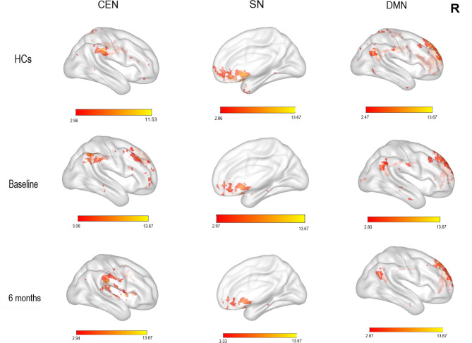
Background: Patients with remitted major depressive disorder (rMDD) show abnormal functional connectivity of the central executive network (CEN), salience networks (SN) and default mode network (DMN). It is unclear how these change during remission, or whether changes are related to function.
Methods: Three spatial networks in 17 patients with rMDD were compared between baseline and the six-month follow-up, and to 22 healthy controls. Correlations between these changes and psychosocial functioning were also assessed.
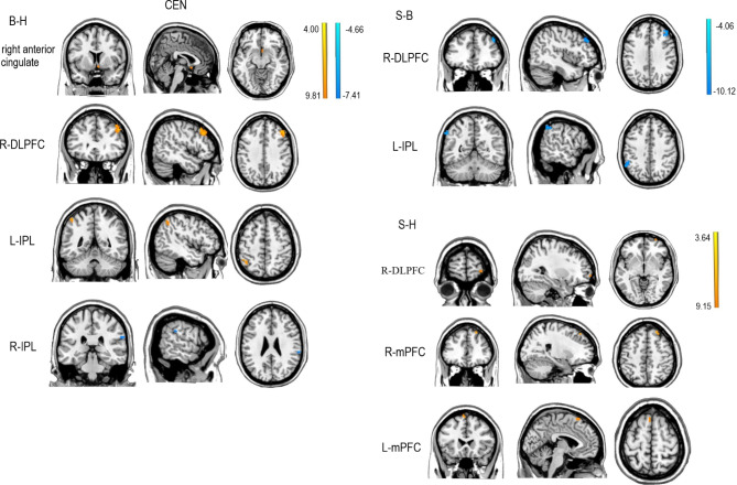
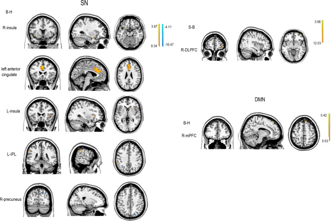
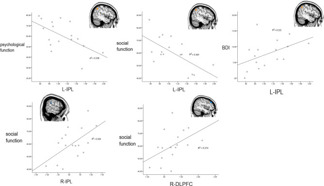
Results: In the CEN, patients at baseline had abnormal functional connectivity in the right anterior cingulate, right dorsolateral prefrontal cortex (DLPFC) and inferior parietal lobule (IPL) compare with HCs. There were functional connection differences in the right DLPFC and left IPL at baseline during follow-up. Abnormal connectivity in the right DLPFC and medial prefrontal cortex (mPFC) were found at follow-up. In the SN, patients at baseline had abnormal functional connectivity in the insula, left anterior cingulate, left IPL, and right precuneus; compared with baseline, patients had higher connectivity in the right DLPFC at follow-up. In the DMN, patients at baseline had abnormal functional connectivity in the right mPFC. Resting-state functional connectivity of the IPL and DLPFC in the CEN correlated with psychosocial functioning.
Conclusions: At six-month follow-up, the CEN still showed abnormal functional connectivity in those with rMDD, while anomalies in the SN and DMN has disappeared. Resting-state functional connectivity of the CEN during early rMDD is associated with psychosocial function.
Clinical trials registration: Pharmacotherapy and Psychotherapy for MDD after Remission on Psychology and Neuroimaging. https://www.
Clinicaltrials: gov/ , registration number: NCT01831440 (15/4/2013).
Keywords: Central executive network; Default mode network; Psychosocial functioning; Remitted major depressive disorder; Salience network.
© 2023. BioMed Central Ltd., part of Springer Nature.
Conflict of interest statement
The authors declare no competing interests.
Figures
Similar articles
-
The alteration of cognitive function networks in remitted patients with major depressive disorder: an independent component analysis.Behav Brain Res. 2021 Feb 26;400:113018. doi: 10.1016/j.bbr.2020.113018. Epub 2020 Dec 7. Behav Brain Res. 2021. PMID: 33301816
-
Functional connectivity profiles in remitted depression and their relation to ruminative thinking.Neuroimage Clin. 2025;45:103716. doi: 10.1016/j.nicl.2024.103716. Epub 2024 Nov 26. Neuroimage Clin. 2025. PMID: 39622113 Free PMC article.
-
Cognitive behavioral therapy may rehabilitate abnormally functional communication pattern among the triple-network in major depressive disorder: A follow-up study.J Affect Disord. 2022 May 1;304:28-39. doi: 10.1016/j.jad.2022.02.050. Epub 2022 Feb 19. J Affect Disord. 2022. PMID: 35192866
-
Resting state brain network function in major depression - Depression symptomatology, antidepressant treatment effects, future research.J Psychiatr Res. 2017 Sep;92:147-159. doi: 10.1016/j.jpsychires.2017.04.007. Epub 2017 Apr 24. J Psychiatr Res. 2017. PMID: 28458140 Review.
-
Dysregulation within the salience network and default mode network in hyperthyroid patients: a follow-up resting-state functional MRI study.Brain Imaging Behav. 2020 Feb;14(1):30-41. doi: 10.1007/s11682-018-9961-6. Brain Imaging Behav. 2020. PMID: 30259292 Review.
Cited by
-
Comparative effects of 10-Hz rTMS and iTBS on cortico-striatal connectivity in major depressive disorder: a sham-controlled study.Mol Psychiatry. 2025 Jun 28. doi: 10.1038/s41380-025-03091-0. Online ahead of print. Mol Psychiatry. 2025. PMID: 40581658
-
Transcutaneous auricular vagus nerve stimulation can modulate fronto-parietal brain networks.Front Neurosci. 2024 Jul 18;18:1368754. doi: 10.3389/fnins.2024.1368754. eCollection 2024. Front Neurosci. 2024. PMID: 39091347 Free PMC article.
-
Underexplored Connections Between Diabetes, Hypomanic States and Insecure Attachment.Psychol Res Behav Manag. 2025 Jun 11;18:1333-1345. doi: 10.2147/PRBM.S524823. eCollection 2025. Psychol Res Behav Manag. 2025. PMID: 40525100 Free PMC article. Review.
References
-
- Ustun TB, Chatterji S. Global burden of depressive disorders and future projections. In: Dawson A, Tylee A, editors. Depression: social and economic timebomb. London: BMJ Books; 2001.
-
- World Health Organization. Depression and other common mental disorders: global health estimates. 2017. http://apps.who.int/iris/bitstream/10665/254610/1/WHO-MSD-MER. Accessed 27 August, 2017.
-
- Frank E, Prien RF, Jarrett RB, Keller MB, Kupfer DJ, Lavori PW, Rush AJ, Weissman MM. Conceptualization and rationale for consensus definitions of terms in major depressive disorder: remission, recovery, relapse, and recurrence. A.M.A. Arch Gen Psychiatry. 1991;48(9):851–5. doi: 10.1001/archpsyc.1991.01810330075011. - DOI - PubMed
Publication types
MeSH terms
Associated data
LinkOut - more resources
Full Text Sources
Medical
