

Diagnostic performance of Strongyloides-specific IgG4 detection in urine for diagnosis of human strongyloidiasis
- PMID: 37641157
- PMCID: PMC10464225
- DOI: 10.1186/s13071-023-05935-6
Diagnostic performance of Strongyloides-specific IgG4 detection in urine for diagnosis of human strongyloidiasis
Abstract
Background: Detection of parasite-specific IgG in urine is a sensitive method for diagnosis of strongyloidiasis and gives similar accuracy to serum IgG. However, there are no data concerning detection of IgG subclass in urine. To further explore the utility of diagnosis from urine samples, we evaluated the diagnostic performance of IgG4 in urine compared with parasitological and other immunological methods.
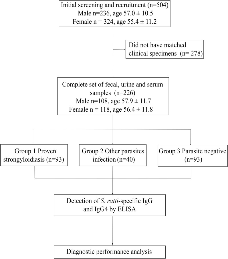
Methods: The urine and sera included proven strongyloidiasis (group 1, n = 93), other parasitic infections (group 2, n = 40) and parasite negatives (group 3, n = 93). The performance of Strongyloides-specific IgG4 in urine for diagnosis of strongyloidiasis using fecal examinations as the reference standard was assessed.
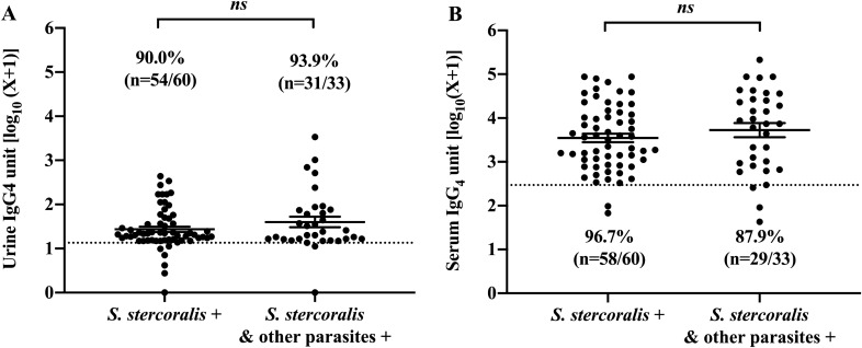
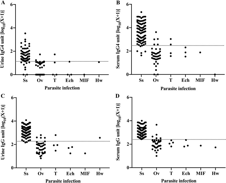
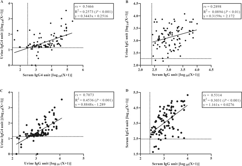
Results: With fecal examination as a gold standard, Strongyloides-specific IgG4 in urine had 91.4% sensitivity and 93.2% specificity while serum IgG4 had 93.6% sensitivity and 91.0% specificity. IgG4 in both urine and serum had almost perfect diagnostic agreements with fecal examination (Cohen's kappa coefficient was > 0.8). Cross-reactivity to Opisthorchis viverrini and Taenia spp. of IgG4 in urine were 7.5% and 12.5% in serum. Concurrent analyses of total IgG in urine and serum showed that the sensitivities (97.9-100%) and specificities (88.7-91.0%) were similar (P > 0.05). The sensitivity for parasitological examination by the formalin-ethyl acetate concentration technique (FECT) was 49.5% and that for agar plate culture technique (APC) it was 92.6%.
Conclusion: Our findings showed that specific IgG4 detection in urine yielded similar diagnostic performance to the same biomarkers in serum. This suggests that accurate diagnosis of strongyloidiasis can be performed using urine samples and IgG4 is a valid choice of diagnostic marker. Further assessment is required to assess the utility of urine IgG4 for measuring the response treatment in strongyloidiasis.
Keywords: Enzyme-linked immunosorbent assay (ELISA); Immunoglobulin G4; Strongyloides stercoralis.
© 2023. BioMed Central Ltd., part of Springer Nature.
Conflict of interest statement
The authors declare that they have no competing interests.
Figures
Similar articles
-
Partially purified Strongyloides ratti antigen improved the diagnostic performance of strongyloidiasis by enzyme-linked immunosorbent assay (ELISA) and immunochromatographic test (ICT).Microbiol Spectr. 2025 Mar 4;13(3):e0236824. doi: 10.1128/spectrum.02368-24. Epub 2025 Feb 4. Microbiol Spectr. 2025. PMID: 39902967 Free PMC article.
-
Analysis of Daily Variation for 3 and for 30 Days of Parasite-Specific IgG in Urine for Diagnosis of Strongyloidiasis by Enzyme-Linked Immunosorbent Assay.Acta Trop. 2021 Jun;218:105896. doi: 10.1016/j.actatropica.2021.105896. Epub 2021 Mar 20. Acta Trop. 2021. PMID: 33753029
-
Diagnostic accuracy of a novel enzyme-linked immunoassay for the detection of IgG and IgG4 against Strongyloides stercoralis based on the recombinant antigens NIE/SsIR.Parasit Vectors. 2021 Aug 18;14(1):412. doi: 10.1186/s13071-021-04916-x. Parasit Vectors. 2021. PMID: 34407876 Free PMC article.
-
Serodiagnosis and early detection of Strongyloides stercoralis infection.J Microbiol Immunol Infect. 2019 Jun;52(3):371-378. doi: 10.1016/j.jmii.2018.10.001. Epub 2018 Oct 11. J Microbiol Immunol Infect. 2019. PMID: 30482708 Review.
-
Metaperiodate deglycosylation of Strongyloides venezuelensis larvae: Immunochemical characterization and antigen production for human strongyloidiasis diagnosis.Acta Trop. 2018 Jun;182:27-33. doi: 10.1016/j.actatropica.2018.02.001. Epub 2018 Feb 15. Acta Trop. 2018. PMID: 29454735 Review.
Cited by
-
Strongyloidiasis.Nat Rev Dis Primers. 2024 Jan 25;10(1):6. doi: 10.1038/s41572-023-00490-x. Nat Rev Dis Primers. 2024. PMID: 38272922 Review.
-
Partially purified Strongyloides ratti antigen improved the diagnostic performance of strongyloidiasis by enzyme-linked immunosorbent assay (ELISA) and immunochromatographic test (ICT).Microbiol Spectr. 2025 Mar 4;13(3):e0236824. doi: 10.1128/spectrum.02368-24. Epub 2025 Feb 4. Microbiol Spectr. 2025. PMID: 39902967 Free PMC article.
References
MeSH terms
Substances
LinkOut - more resources
Full Text Sources
Miscellaneous
