Intra- and interobserver reproducibility of transvaginal ultrasound for the detection and measurement of endometriotic lesions of the bowel
- PMID: 37641421
- PMCID: PMC10541154
- DOI: 10.1111/aogs.14660
Intra- and interobserver reproducibility of transvaginal ultrasound for the detection and measurement of endometriotic lesions of the bowel
Abstract
Introduction: The number and invasion depth of endometriotic bowel lesions, total length of bowel affected by endometriosis, lesion-to-anal verge distance, and extent of pouch of Douglas obliteration are important factors in preoperatively determining risk and complexity of endometriosis surgery. The intra- and interobserver reproducibility of transvaginal ultrasound in the evaluation of many of these parameters has not yet been investigated. Our study aimed to assess the intra- and interobserver reproducibility of transvaginal ultrasound between an experienced and less experienced examiner for all of these parameters.
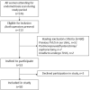
Material and methods: This prospective observational cross-sectional study was conducted between July 2019 and November 2020. Fifty consecutive premenopausal women who underwent transvaginal ultrasound examination in our clinic for the first time, were examined by the same two operators during the same attendance. Outcomes of interest were the inter-rater reproducibility of transvaginal ultrasound for detecting the presence, number, depth and size of bowel endometriotic nodules, lesion-to-anal-verge distance, total length of bowel affected, and pouch of Douglas obliteration. The intraobserver reproducibility was assessed for the continuous parameters. Cohen's kappa (κ) statistic, Cohen's weighted kappa (κ), proportions of agreement, intraclass correlation coefficient (ICC) and Bland-Altman limits of agreement were used to assess the reproducibility of the parameters.
Results: The inter-rater agreement and reliability were very good for identifying bowel endometriosis, the number and invasion depth of bowel nodules, determining whether the maximum nodule length was <3 cm, and lesion-to-anal-verge distance <8 cm (proportion of agreement 0.92, 0.94, 0.97, 0.94, 0.96; κ 0.92, 0.91, 0.92, 0.82, 0.89). The inter-rater agreement and reliability were good for assessing pouch of Douglas obliteration (proportion of agreement 0.86, κ 0.80). The intra-rater reliability for the mean nodule diameter (ICC 0.93 and 0.97) and total length of bowel affected (ICC 0.94 and 0.91) were excellent for operators A and B, respectively. The inter-rater reliability for the mean nodule diameter was good (ICC 0.80), and moderate for the total length of bowel affected (ICC 0.70). The Bland-Altman limits of agreement demonstrated clinically acceptable ranges for these two parameters.
Conclusions: This study demonstrated a high intra- and inter-rater reproducibility of transvaginal ultrasound in the diagnosis of bowel endometriosis and measurement of its various components.
Keywords: bowel endometriosis; deep endometriosis; rectosigmoid colon; reliability; reproducibility; surgery planning; transvaginal ultrasound.
© 2023 The Authors. Acta Obstetricia et Gynecologica Scandinavica published by John Wiley & Sons Ltd on behalf of Nordic Federation of Societies of Obstetrics and Gynecology (NFOG).
Conflict of interest statement
TT reports receiving personal fees for lectures on ultrasound from GE Healthcare, Samsung, Medtronic and Merck, outside of this study. The other authors report no conflict of interest.
Figures


Similar articles
-
Intra- and interobserver reproducibility of pelvic ultrasound for the detection and measurement of endometriotic lesions.Hum Reprod Open. 2020 Mar 6;2020(2):hoaa001. doi: 10.1093/hropen/hoaa001. eCollection 2020. Hum Reprod Open. 2020. PMID: 32161818 Free PMC article.
-
The prediction of pouch of Douglas obliteration using offline analysis of the transvaginal ultrasound 'sliding sign' technique: inter- and intra-observer reproducibility.Hum Reprod. 2013 May;28(5):1237-46. doi: 10.1093/humrep/det044. Epub 2013 Mar 12. Hum Reprod. 2013. PMID: 23482338
-
Transvaginal sonography accurately measures lesion-to-anal-verge distance in women with deep endometriosis of the rectosigmoid.Ultrasound Obstet Gynecol. 2020 Nov;56(5):766-772. doi: 10.1002/uog.21995. Ultrasound Obstet Gynecol. 2020. PMID: 32068921
-
Current Status of Transvaginal Ultrasound Accuracy in the Diagnosis of Deep Infiltrating Endometriosis Before Surgery: A Systematic Review of the Literature.J Ultrasound Med. 2020 Aug;39(8):1477-1490. doi: 10.1002/jum.15246. Epub 2020 Feb 21. J Ultrasound Med. 2020. PMID: 32083336
-
Ultrasonography for bowel endometriosis.Best Pract Res Clin Obstet Gynaecol. 2021 Mar;71:38-50. doi: 10.1016/j.bpobgyn.2020.05.010. Epub 2020 Jun 6. Best Pract Res Clin Obstet Gynaecol. 2021. PMID: 32620408 Review.
Cited by
-
Development of deep pelvic endometriosis following acute haemoperitoneum: a prospective ultrasound study.Hum Reprod Open. 2024 May 29;2024(3):hoae036. doi: 10.1093/hropen/hoae036. eCollection 2024. Hum Reprod Open. 2024. PMID: 38905001 Free PMC article.
-
Ultrasound of the uterosacral ligaments: A reliability study for diagnosing endometriosis in Australian non-specialised medical imaging and radiology settings.Australas J Ultrasound Med. 2025 Feb 18;28(1):e12420. doi: 10.1002/ajum.12420. eCollection 2025 Feb. Australas J Ultrasound Med. 2025. PMID: 39975473
-
Understanding and addressing female pelvic pain - a multifaceted challenge.Acta Obstet Gynecol Scand. 2023 Oct;102(10):1248-1249. doi: 10.1111/aogs.14682. Acta Obstet Gynecol Scand. 2023. PMID: 37773602 Free PMC article. No abstract available.
References
-
- Bean E, Naftalin J, Horne A, Saridogan E, Cutner A, Jurkovic D. Prevalence of deep and ovarian endometriosis in early pregnancy: ultrasound diagnostic study. Ultrasound Obstet Gynecol. 2021;59:107‐113. - PubMed
-
- Abrao MS, Petraglia F, Falcone T, Keckstein J, Osuga Y, Chapron C. Deep endometriosis infiltrating the recto‐sigmoid:critical factors to consider before management. Hum Reprod Open. 2015;21:329‐339. - PubMed
-
- Nezhat C, Li A, Falik R, et al. Bowel endometriosis: diagnosis and management. AJOG. 2017;218:549‐562. - PubMed
-
- Keckstein J, Saridogan E, Ulrich UA, et al. The #Enzian classification: a comprehensive non‐invasive and surgical description system for endometriosis. Acta Obstet Gynecol Scan. 2021;100:1165‐1175. - PubMed
Publication types
MeSH terms
LinkOut - more resources
Full Text Sources
Medical

