

Optic nerve head perfusion changes in eyes with proliferative diabetic retinopathy treated with intravitreal ranibizumab or photocoagulation: a randomized controlled trial
- PMID: 37641606
- PMCID: PMC10460245
- DOI: 10.51329/mehdiophthal1459
Optic nerve head perfusion changes in eyes with proliferative diabetic retinopathy treated with intravitreal ranibizumab or photocoagulation: a randomized controlled trial
Abstract
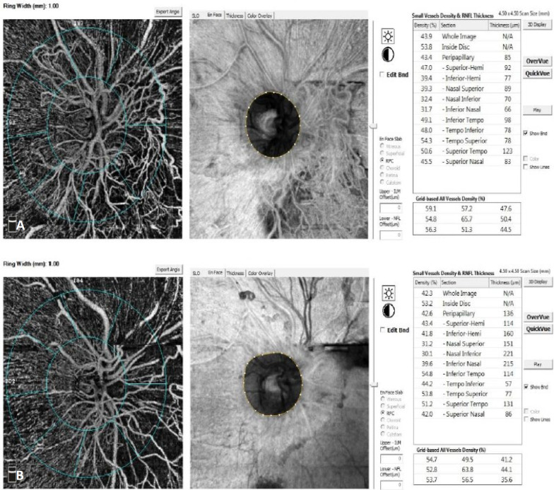
Background: Proliferative diabetic retinopathy (PDR) is a serious sight-threatening disease, and half of the patients with high-risk PDR can develop legal blindness within 5 years, if left untreated. This study was aimed at comparing panretinal photocoagulation (PRP) and intravitreal ranibizumab injections in terms of radial peripapillary capillary (RPC) density on optical coherence tomography angiography (OCTA) in patients with treatment-naive PDR.
Methods: This open-label, prospective, randomized clinical trial included 50 patients with treatment-naive PDR with optic disc neovascularization and randomized them into two groups: group 1, with patients undergoing two sessions of PRP 2 weeks apart, and group 2, with patients received three intravitreal ranibizumab injections (0.5 mg) 1 month apart for 3 consecutive months. Patients underwent a full ophthalmological examination, including best-corrected distance visual acuity (BCDVA) measurement in the logarithm of minimal angle of resolution (logMAR) notation and OCTA before intervention and monthly after the last laser session or the first intravitreal ranibizumab injection for 3 months of follow-up. Visual field (VF) was tested at the beginning and end of 3 months.
Results: Forty-two (84%) eyes completed the 3-month follow-up, including 22 eyes in the PRP group (88%) and 20 (80%) eyes in the ranibizumab group. The two groups were comparable in terms of demographic characteristics, diabetes duration, baseline BCDVA, glycated hemoglobin level, OCTA parameters, VF indices, and intraocular pressure (all P > 0.05). The RPC density change from baseline to the 3-month follow-up was significantly lower in the PRP group than in the ranibizumab group (mean difference in RPC density change: - 3.61%; 95% confidence interval: - 5.57% to - 1.60%; P = 0.001). The median (interquartile range) logMAR change from baseline to the 3-month follow-up (0.0 [0.2]) was significantly higher in the PRP group than in the ranibizumab group (- 0.15 [0.3]; P < 0.05). The median changes in central foveal thickness from baseline to the 3-month follow-up differed significantly between the two groups (P = 0.001).
Conclusions: In eyes with PDR and neovascularization of the disc RPC density on OCTA increased in the ranibizumab group and decreased in the PRP group. Visual acuity gain was higher in the ranibizumab group than in the PRP group. Future multicenter trials addressing our limitations are required to verify the findings of this study.
Keywords: diabetic retinopathies; intravitreal injection; laser ablation; laser therapies; lucentis; optic nerves; optical coherence tomography; photocoagulation.
© Author(s).
Conflict of interest statement
None.
Figures


Similar articles
-
Panretinal photocoagulation (PRP) versus PRP plus intravitreal ranibizumab for high-risk proliferative diabetic retinopathy.Acta Ophthalmol. 2011 Nov;89(7):e567-72. doi: 10.1111/j.1755-3768.2011.02184.x. Epub 2011 Jul 5. Acta Ophthalmol. 2011. PMID: 21726427
-
Factors Associated with Worsening Proliferative Diabetic Retinopathy in Eyes Treated with Panretinal Photocoagulation or Ranibizumab.Ophthalmology. 2017 Apr;124(4):431-439. doi: 10.1016/j.ophtha.2016.12.005. Epub 2017 Feb 1. Ophthalmology. 2017. PMID: 28161147 Free PMC article. Clinical Trial.
-
Panretinal photocoagulation versus panretinal photocoagulation plus intravitreal bevacizumab for high-risk proliferative diabetic retinopathy.Int J Ophthalmol. 2016 Dec 18;9(12):1772-1778. doi: 10.18240/ijo.2016.12.12. eCollection 2016. Int J Ophthalmol. 2016. PMID: 28003978 Free PMC article.
-
Intravitreal steroids for macular edema in diabetes.Cochrane Database Syst Rev. 2020 Nov 17;11(11):CD005656. doi: 10.1002/14651858.CD005656.pub3. Cochrane Database Syst Rev. 2020. PMID: 33206392 Free PMC article.
-
Practical Lessons from Protocol I for the Management of Diabetic Macular Edema.Dev Ophthalmol. 2017;60:91-108. doi: 10.1159/000459692. Epub 2017 Apr 20. Dev Ophthalmol. 2017. PMID: 28427069 Review.
Cited by
-
Effect of panretinal photocoagulation versus intravitreal bevacizumab injection on optic disc microcirculation in patients with diabetic retinopathy.Int J Retina Vitreous. 2024 Dec 18;10(1):98. doi: 10.1186/s40942-024-00621-w. Int J Retina Vitreous. 2024. PMID: 39695775 Free PMC article.
-
Effects of anti‑VEGF on peripapillary retinal nerve fiber layer and papillary/peripapillary blood circulation in retinopathies (Review).Int J Mol Med. 2025 Sep;56(3):133. doi: 10.3892/ijmm.2025.5574. Epub 2025 Jul 4. Int J Mol Med. 2025. PMID: 40613237 Free PMC article. Review.
References
-
- Gross JG, Glassman AR, Jampol LM, Inusah S, Aiello LP, Antoszyk AN, et al. Writing Committee for the Diabetic Retinopathy Clinical Research Network. Panretinal Photocoagulation vs Intravitreous Ranibizumab for Proliferative Diabetic Retinopathy: A Randomized Clinical Trial. JAMA. 2015;314(20):2137–2146. - PMC - PubMed
-
- Reddy SV, Husain D. Panretinal Photocoagulation: A Review of Complications. Semin Ophthalmol. 2018;33(1):83–88. - PubMed
-
- Fong DS, Girach A, Boney A. Visual side effects of successful scatter laser photocoagulation surgery for proliferative diabetic retinopathy: a literature review. Retina. 2007;27(7):816–24. - PubMed
-
- Heidary F, Hitam WH, Ngah NF, George TM, Hashim H, Shatriah I. Intravitreal Ranibizumab for Choroidal Neovascularization in Best’s Vitelliform Macular Dystrophy in a 6-Year-Old Boy. J Pediatr Ophthalmol Strabismus. 2011;48 Online:e19–22. - PubMed
LinkOut - more resources
Full Text Sources
Research Materials