Review
. 2023 Aug 23:16:19-41.
doi: 10.2147/SCCAA.S417842. eCollection 2023.

Mitochondria in Cancer Stem Cells: From an Innocent Bystander to a Central Player in Therapy Resistance

Affiliations
Review

Mitochondria in Cancer Stem Cells: From an Innocent Bystander to a Central Player in Therapy Resistance

Sireesha V Garimella et al. Stem Cells Cloning. .

Abstract

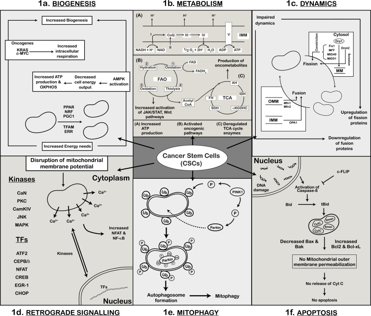
Cancer continues to rank among the world's leading causes of mortality despite advancements in treatment. Cancer stem cells, which can self-renew, are present in low abundance and contribute significantly to tumor recurrence, tumorigenicity, and drug resistance to various therapies. The drug resistance observed in cancer stem cells is attributed to several factors, such as cellular quiescence, dormancy, elevated aldehyde dehydrogenase activity, apoptosis evasion mechanisms, high expression of drug efflux pumps, protective vascular niche, enhanced DNA damage response, scavenging of reactive oxygen species, hypoxic stability, and stemness-related signaling pathways. Multiple studies have shown that mitochondria play a pivotal role in conferring drug resistance to cancer stem cells, through mitochondrial biogenesis, metabolism, and dynamics. A better understanding of how mitochondria contribute to tumorigenesis, heterogeneity, and drug resistance could lead to the development of innovative cancer treatments.

Keywords: cancer stem cells; drug resistance; metabolic dysfunction; mitochondria; therapy.


Conflict of interest statement

The authors declare no conflicts of interest in this work.

Figures

Figure 1
Figure 1
Mitochondrial Dysfunction and Cancer Stem Cells (CSCs). (a) Schematic of mitochondrial biogenesis and its regulation by transcription factors (PGC1, NRF, TFAM, PPAR, and ERR). Additionally, depicted are AMPK, oncogenic KRAS, and c-MYC-dependent mechanisms that lead to increase in biogenesis and energy production. This results in elevated oxidative phosphorylation (OXPHOS) and high ATP levels in CSCs. (b) Representation of mitochondrial metabolic dependency in CSCs. Cellular energy is derived through OXPHOS, fatty acid oxidation (FAO), and the TCA cycle within the mitochondria. CSCs exhibit increased oxidative phosphorylation for enhanced ATP production and elevated fatty acid oxidation through activation of oncogenic pathways. Deregulated TCA cycle enzymes in CSCs produce oncometabolites contributing to cancer progression. (c) Representation of altered mitochondrial dynamics in CSCs, where the balance between mitochondrial fission and fusion is disrupted. Upregulation of mitochondrial fission proteins (Drp1) and their regulators (Fis1, MID49, MID51, MFF) and downregulation of mitochondrial fusion proteins (Mfn1, Mfn2, OPA1) leads to impaired mitochondrial dynamics. (d) Increased activity of Ca2+-dependent kinases (PKC, CaN, CAMKIV, JNK, MAPK) due to altered membrane potential in CSCs is shown. Also indicated are the kinases and nuclear transcription factors involved in retrograde signaling. (e) Schematic representation of mitophagy in CSCs. Elevated cytoplasmic PINK1 phosphorylates Parkin and ubiquitinated-OMM proteins. Phosphorylated Parkin is transported into the mitochondria where it ubiquitinates itself and other mitochondrial substrates. These ubiquitin (Ub)-marked mitochondria are degraded by autophagosomes. (f) Mitochondria-mediated apoptosis in CSCs. Cells with damaged DNA activate caspase-8 mediated cell death. In CSCs, activation of caspase-8 is inhibited by high levels of c-FLIP; levels of pro-apoptotic proteins (Bax, Bak) are decreased while levels of anti-apoptotic proteins (Bcl-xL) are increased leading to cell survival and no apoptosis.

Similar articles

Cited by

References

    1. Ebben JD, Treisman DM, Zorniak M, Kutty RG, Clark PA, Kuo JS. The cancer stem cell paradigm: a new understanding of tumor development and treatment. Expert Opin Ther Targets. 2010;14(6):621–632. PMID: 20426697. doi:10.1517/14712598.2010.485186 - DOI - PMC - PubMed
    1. Lapidot T, Sirard C, Vormoor J, et al. A cell initiating human acute myeloid leukaemia after transplantation into SCID mice. Nature. 1994;367:645–648. PMID: 7509044. doi:10.1038/367645a0 - DOI - PubMed
    1. Kleinsmith LJ, Pierce GB. Multipotentiality of Single Embryonal Carcinoma Cells. Cancer Res. 1964;24:1544–1551. PMID: 14234000. - PubMed
    1. Bonnet D, Dick JE. Human acute myeloid leukemia is organized as a hierarchy that originates from a primitive hematopoietic cell. Nat Med. 1997;3:730–737. PMID: 9212098. doi:10.1038/nm0797-730 - DOI - PubMed
    1. Makino S. Further evidence favoring the concept of the stem cell in ascites tumors of rats. Ann N Y Acad Sci. 1956;63:818–830. PMID: 13314436. doi:10.1111/j.1749-6632.1956.tb50894.x - DOI - PubMed