. 2023 Sep 1;27(5):493-511.
doi: 10.4196/kjpp.2023.27.5.493.

Inhibition of the interaction between Hippo/YAP and Akt signaling with ursolic acid and 3'3-diindolylmethane suppresses esophageal cancer tumorigenesis

Affiliations

Inhibition of the interaction between Hippo/YAP and Akt signaling with ursolic acid and 3'3-diindolylmethane suppresses esophageal cancer tumorigenesis

Ruo Yu Meng et al. Korean J Physiol Pharmacol. .

Abstract

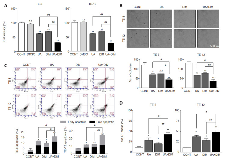
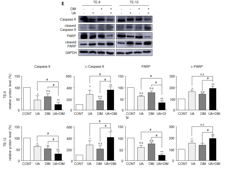
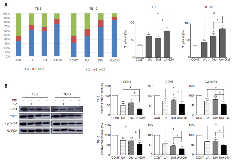
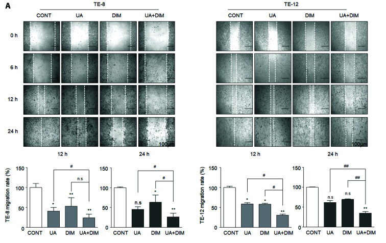
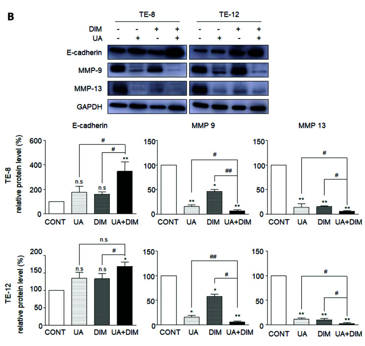
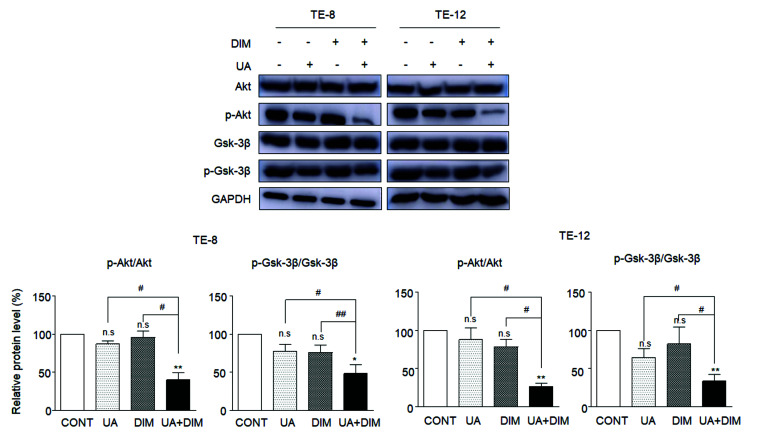
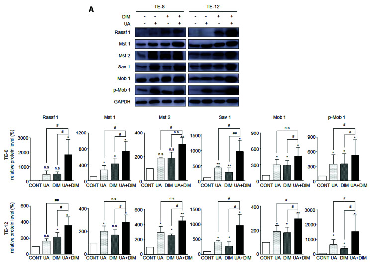
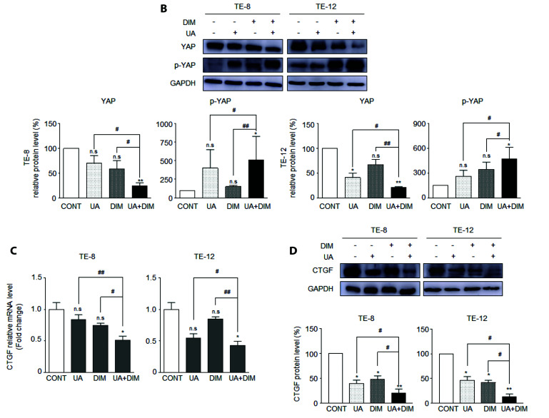
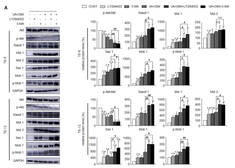
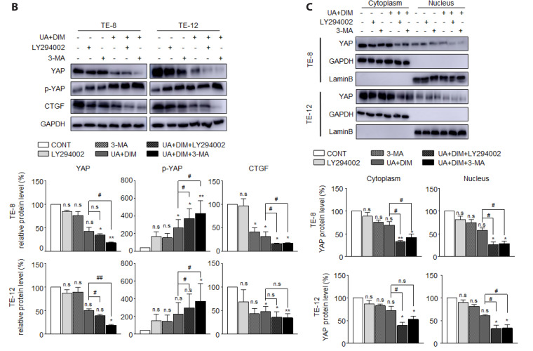
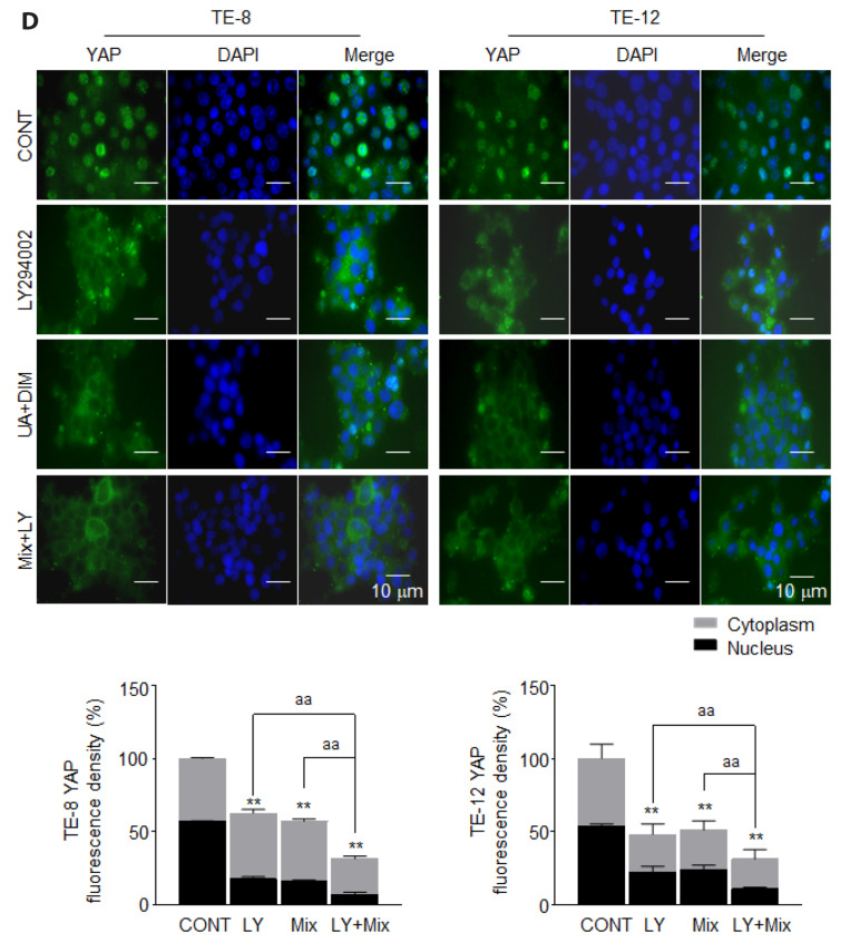
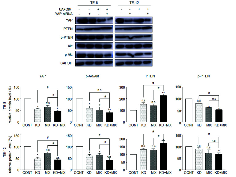
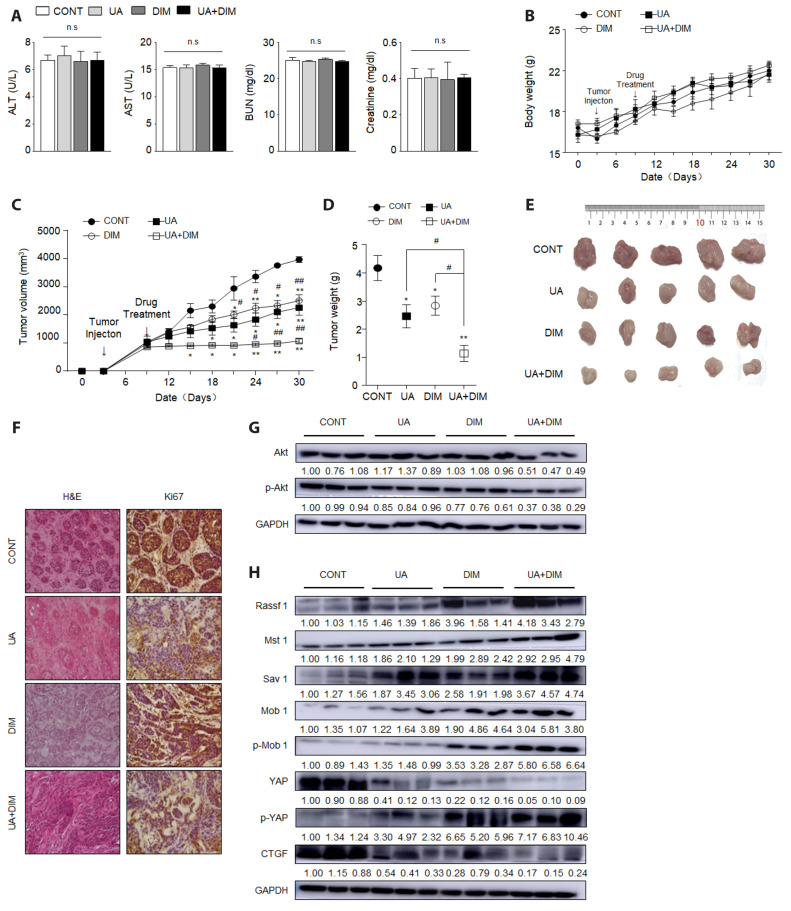
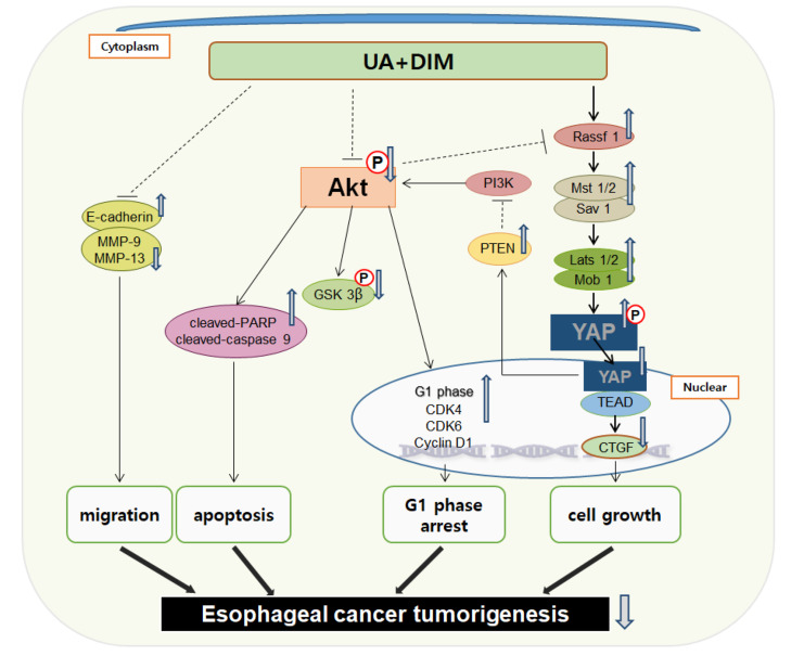
Hippo/YAP signaling hinders cancer progression. Inactivation of this pathway contributes to the development of esophageal cancer by activation of Akt. However, the possible interaction between Akt and Hippo/YAP pathways in esophageal cancer progression is unclear. In this study, we found that ursolic acid (UA) plus 3'3-diindolylmethane (DIM) efficiently suppressed the oncogenic Akt/Gsk-3β signaling pathway while activating the Hippo tumor suppressor pathway in esophageal cancer cells. Moreover, the addition of the Akt inhibitor LY294002 and the PI3K inhibitor 3-methyladenine enhanced the inhibitory effects of UA plus DIM on Akt pathway activation and further stimulated the Hippo pathway, including the suppression of YAP nuclear translocation in esophageal cancer cells. Silencing YAP under UA plus DIM conditions significantly increased the activation of the tumor suppressor PTEN in esophageal cancer cells, while decreasing p-Akt activation, indicating that the Akt signaling pathway could be down-regulated in esophageal cancer cells by targeting PTEN. Furthermore, in a xenograft nude mice model, UA plus DIM treatment effectively diminished esophageal tumors by inactivating the Akt pathway and stimulating the Hippo signaling pathway. Thus, our study highlights a feedback loop between the PI3K/Akt and Hippo signaling pathways in esophageal cancer cells, implying that a low dose of UA plus DIM could serve as a promising chemotherapeutic combination strategy in the treatment of esophageal cancer.

Keywords: 3′,3-diindolylmethane; Akt; Esophageal squamous cancer cells; Hippo signaling pathway; Ursolic acid; YAP.


Conflict of interest statement

CONFLICTS OF INTEREST

The authors declare no conflicts of interest.

Figures

Fig. 1
Fig. 1. Ursolic acid (UA) plus 3,3’-diindolylmethane (DIM) inhibits cell growth and promotes apoptosis in ESCC.
(A) UA and DIM combination therapy inhibited cell viability in TE-8 and TE-12 cells. Cells were treated with UA (20 μM), DIM (50 μM), and combination treatment for 48 h. An EZ-CYTOX assay kit was used to assess the cell viability of esophageal cancer cells and to generate growth curves. (B) UA plus DIM combination reduced colony number and size in TE-8 and TE-12 cells. Colonies were cultured with UA (20 μM), DIM (50 μM), and the combination treatment of UA and DIM for 4 weeks in soft agar gel. The quantification of colony numbers was calculated by ImageJ. (C) UA plus DIM increased early-stage and late-stage apoptosis in TE-8 and TE-12 cells. Annexin V/PI staining was performed to detect cell apoptosis by flow cytometry. (D) UA plus DIM combination induced subG1 phase cell accumulation in TE-8 and TE-12 cells. Cells were treated with UA (20 μM), DIM (50 μM), and combination treatment for 48 h. SubG1 phase cells were indicated by apoptosis markers and detected by flow cytometry. (E) UA plus DIM enhanced apoptosis-related protein levels. Expression of apoptosis-regulatory factor proteins caspase-9/cleaved-caspase-9 and PRAP/cleaved-PARP were examined by Western blot analysis. The quantification of protein bands was estimated by ImageJ software. GAPDH was used as the internal control. Data are expressed as the mean ± standard error of the mean. CONT, control; DMSO, dimethyl sulfoxide; ESCC, esophageal squamous cell carcinoma; PI, propidium iodide; n.s., no significant. *, compared to the control; #, compare with UA plus DIM combination treatment. * or #, p < 0.05; ** or ##, p < 0.01.
Fig. 2
Fig. 2. Ursolic acid (UA) plus 3,3’-diindolylmethane (DIM) induces G1 phase arrest in ESCC.
(A) UA plus DIM induced G1 phase arrest in TE-8 and TE-12 cells. Cells were treated with UA (20 μM), DIM (50 μM), and a combination of both for 48 h. PI staining-based flow cytometry assay was designed to study the cell cycle in esophageal cancer cells. (B) UA plus DIM combination suppressed G1 phase-related protein levels. Western blot analysis was performed to measure G1 phase-related protein CDK4, CDK6, and cyclin D1. GAPDH was used as the internal control. Data are expressed as the mean ± standard error of the mean. CONT, control; ESCC, esophageal squamous cell carcinoma; PI, propidium iodide; n.s., no significant. *, compared to the control; #, compare with UA plus DIM combination treatment. * or #, p < 0.05; ** or ##, p < 0.01.
Fig. 3
Fig. 3. Ursolic acid (UA) plus 3,3’-diindolylmethane (DIM) suppresses cell migration in ESCC.
(A) UA (20 μM) and DIM (50 μM) combination treatment inhibited cell migration in the presence of 5 µg/ml mitomycin-C. Wound healing assays were used to analyze cell migration. TE-8 and TE-12 cell migration rates were measured at 12 h and 24 h after UA or DIM single treatment or a combination and calculated by ImageJ software. (B) UA plus DIM inhibited migration-related protein expression. Migration-related proteins E-cadherin, MMP-13, and MMP-9 were examined by Western blot analysis. GAPDH was used as the internal control. Data are expressed as the mean ± standard error of the mean. CONT, control; ESCC, esophageal squamous cell carcinoma; n.s., no significant. *, compared to the control; #, compare with UA plus DIM combination treatment. * or #, p < 0.05; ** or ##, p < 0.01.
Fig. 4
Fig. 4. Ursolic acid (UA) plus 3,3’-diindolylmethane (DIM) inactivates the PI3K/Akt/GSK-3β signaling pathway in ESCC.
After TE-8 and TE-12 cells were treated with UA (20 μM), DIM (50 μM), and a combination for 48 h. Akt/p-Akt and Gsk-3β/p-GSK 3β protein levels were examined by Western blot analysis. The quantification of protein bands was estimated by ImageJ software. GAPDH was used as the internal control. Data are expressed as the mean ± standard error of the mean. CONT, control; ESCC, esophageal squamous cell carcinoma; n.s., no significant. *, compared to the control; #, compare with UA plus DIM combination treatment. * or #, p < 0.05; ** or ##, p < 0.01.
Fig. 5
Fig. 5. Ursolic acid (UA) plus 3,3’-diindolylmethane (DIM) actives the Hippo signaling pathway and down-regulates the target gene CTGF in ESCC.
TE-8 and TE-12 cells were treated with UA (20 μM), DIM (50 μM), or combination treatment for 48 h. Western blot analysis was used to determine the Hippo signaling pathway: (A) Rassf1, Mst1, Mst2, Sav1, Mob-1, and p-Mob1, (B) YAP and p-YAP. (C) The Hippo signaling pathway CTGF mRNA level was detected by real-time PCR and (D) protein levels were examined by Western blot analysis. GAPDH was used as the internal control. Data are expressed as the mean ± standard error of the mean. CONT, control; ESCC, esophageal squamous cell carcinoma; n.s., no significant. *, compared to the control; #, compare with UA plus DIM combination treatment. * or #, p < 0.05; ** or ##, p < 0.01.
Fig. 6
Fig. 6. Inhibition of the PI3K/Akt pathway stimulated by ursolic acid (UA) plus 3,3’-diindolylmethane (DIM) combination treatment induces Hippo signaling pathway activation in ESCC.
TE-8 and TE-12 cells were pre-treated with LY294002 (50 μM) or 3-MA (5 mM) for 2 h and further treated with UA (20 μM) plus DIM (50 μM) for 48 h. The expression of Hippo pathway-related proteins: (A) Rassf1, Mst1, Mst2, Sav1, Mob-1, p-Mob1, (B) YAP, and p-YAP and Hippo signaling pathway downstream proteins CTGF were examined by Western blotting. (C) Cytoplasm and nuclear proteins were separated to detect YAP distribution by Western blotting. GAPDH was used as the internal control and cytoplasm marker. Lamin B was used as a nuclear marker. (D) TE-8 and TE-12 cells were stained with anti-YAP antibody (green) and DAPI (blue) and visualized under fluorescence microscopy. Nuclei were stained by DAPI. The value of immunofluorescence was measured and calculated by ImageJ software. Data are expressed as the mean ± standard error of the mean. CONT, control; LY, LY294002; Mix, UA plus DIM; ESCC, esophageal squamous cell carcinoma; n.s., no significant. *, compared to the control; a, combined therapy (mix) compared with mix plus LY294002; #, mix compared with mix plus 3-MA. * or #, p < 0.05; **, ## or aa, p < 0.01.
Fig. 7
Fig. 7. Knockdown of YAP accelerates ursolic acid (UA) plus 3,3’-diindolylmethane (DIM) combination treatment induced down-regulation of the PI3K/Akt pathway by targeting PTEN protein expression in ESCC.
Protein levels of Akt, p-Akt, PTEN, and p-PTEN were measured by Western blot analysis after YAP knockdown by siRNA 24 h following treatment with UA (20 μM) and DIM (50 μM) for 48 h. GAPDH was used as the internal control. Data are expressed as the mean ± standard error of the mean. CONT, control; KD, YAP siRNA knockdown; Mix, UA plus DIM; ESCC, esophageal squamous cell carcinoma; n.s., no significant. *, compared to the control; #, compare with mix plus YAP siRNA knockdown. * or #, p < 0.05; **p < 0.01; ***p < 0.001.
Fig. 8
Fig. 8. Ursolic acid (UA) plus 3,3’-diindolylmethane (DIM) safely and effectively inhibits tumorigenesis through the PI3K/Akt signaling pathway and Hippo signaling pathway in a xenograft mouse model.
TE-8 cells were subcutaneously injected into nude mice to create the xenograft mouse model. After tumors were established, mice were injected with UA, DIM, or combination treatment for 3 weeks. (A) Serum was extracted from mouse blood at 4°C. ALT, AST, BUN, and creatine were used as serum biochemical indices for drug safety evaluation. (B) Body weight and (C) tumor volume was measured every 3 days. (D) Tumor weights were measured after mice were sacrificed. (E) Tumor images were obtained after they were removed from mice. (F) H&E staining (left) and Ki67 staining (right) of tumor tissue (50 uM). Akt-related proteins (G) and Hippo pathway-related proteins (H) are analyzed by Western blotting. Data are expressed as the mean ± standard error of the mean. GAPDH was used as the internal control. CONT, control; ALT, alanine aminotransferase; AST, aspartate aminotransferase; BUN, blood urea nitrogen; n.s., no significant. *, compared to the control; #, compare with UA plus DIM combination treatment. * or #, p < 0.05; ** or ##, p < 0.01.
Fig. 9
Fig. 9. Schematic diagram.
Ursolic acid (UA) plus 3,3’-diindolylmethane (DIM) combination treatment can induce apoptosis by promoting cleaved-PARP and caspase-9, increasing the G1 phase by upregulating CDK4, CDK6, and Cyclin D1 protein expression, and inhibiting migration by regulating E-cadherin, MMP-9, and MMP-13 in esophageal squamous cell carcinoma (ESCC). UA plus DIM combination treatment lowered p-Akt and p-Gsk-3β expression in ESCC. UA plus DIM combination activated the Hippo signaling pathway leading to the inhibition of YAP de-phosphorylation and nuclear translocation and suppression of its downstream gene CTGF further restrained cell growth in ESCC. UA and DIM combination treatment induced the Hippo pathway activation by inhibiting the PI3K/Akt pathway in ESCC. UA plus DIM combination treatment can induce YAP inhibition by conversely inhibiting the PI3K/Akt pathway through up-regulated PTEN. UA plus DIM stimulated a feedback loop between the PI3K/Akt signaling pathway and Hippo signaling pathways in ESCC.

Similar articles

Cited by

References

    1. Uhlenhopp DJ, Then EO, Sunkara T, Gaduputi V. Epidemiology of esophageal cancer: update in global trends, etiology and risk factors. Clin J Gastroenterol. 2020;13:1010–1021. doi: 10.1007/s12328-020-01237-x. - DOI - PubMed
    1. Then EO, Lopez M, Saleem S, Gayam V, Sunkara T, Culliford A, Gaduputi V. Esophageal cancer: an updated surveillance epidemiology and end results database analysis. World J Oncol. 2020;11:55–64. doi: 10.14740/wjon1254. - DOI - PMC - PubMed
    1. Shin A, Won YJ, Jung HK, Kong HJ, Jung KW, Oh CM, Choe S, Lee J. Trends in incidence and survival of esophageal cancer in Korea: analysis of the Korea Central Cancer Registry Database. J Gastroenterol Hepatol. 2018;33:1961–1968. doi: 10.1111/jgh.14289. - DOI - PMC - PubMed
    1. Sun D, Cao M, Li H, He S, Chen W. Cancer burden and trends in China: a review and comparison with Japan and South Korea. Chin J Cancer Res. 2020;32:129–139. doi: 10.21147/j.issn.1000-9604.2020.02.01. - DOI - PMC - PubMed
    1. Cummings D, Wong J, Palm R, Hoffe S, Almhanna K, Vignesh S. Epidemiology, diagnosis, staging and multimodal therapy of esophageal and gastric tumors. Cancers (Basel) 2021;13:582. doi: 10.3390/cancers13030582.f2dd64f9294344139bdb6b4ab170b9c1 - DOI - PMC - PubMed