. 2024 Jan;37(1):50-60.
doi: 10.5713/ab.23.0175. Epub 2023 Aug 28.

Testicular fat deposition attenuates reproductive performance via decreased follicle-stimulating hormone level and sperm meiosis and testosterone synthesis in mouse

Affiliations

Testicular fat deposition attenuates reproductive performance via decreased follicle-stimulating hormone level and sperm meiosis and testosterone synthesis in mouse

Miao Du et al. Anim Biosci. 2024 Jan.

Abstract

Objective: Testicular fat deposition has been reported to affect animal reproduction. However, the underlying mechanism remains poorly understood. The present study explored whether sperm meiosis and testosterone synthesis contribute to mouse testicular fat depositioninduced reproductive performance.

Methods: High fat diet (HFD)-induced obesity CD1 mice (DIO) were used as a testicular fat deposition model. The serum hormone test was performed by agent kit. The quality of sperm was assessed using a Sperm Class Analyzer. Testicular tissue morphology was analyzed by histochemical methods. The expression of spermatocyte marker molecules was monitored by an immuno-fluorescence microscope during meiosis. Analysis of the synthesis of testosterone was performed by real-time polymerase chain reaction and reagent kit.

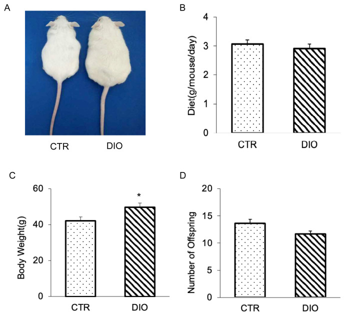
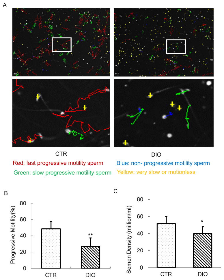
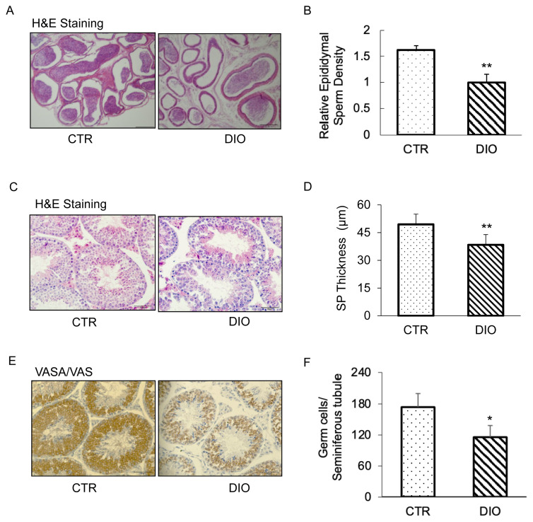
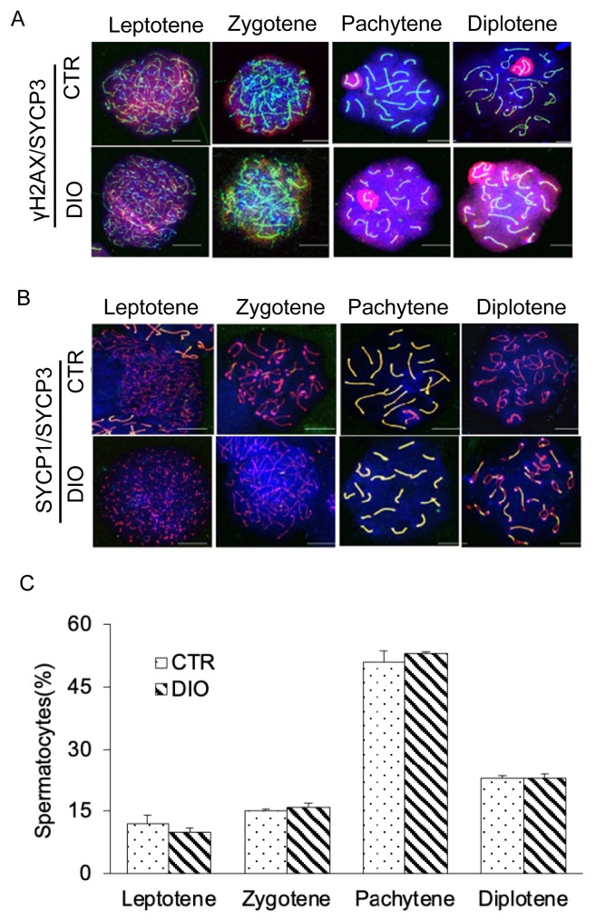
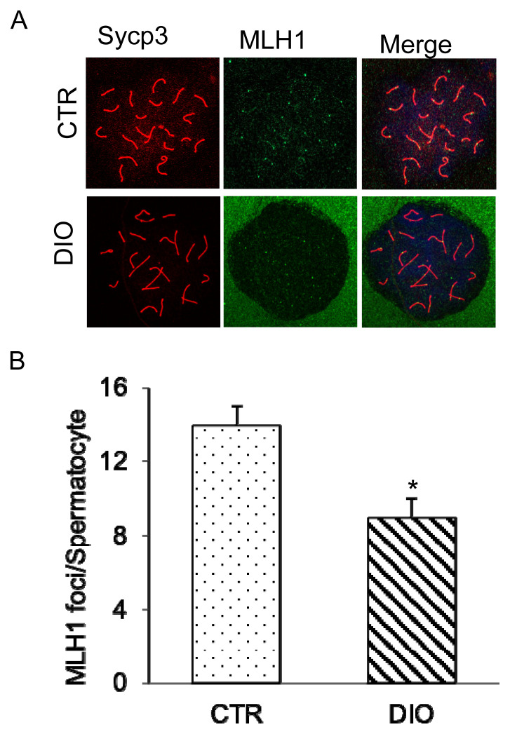
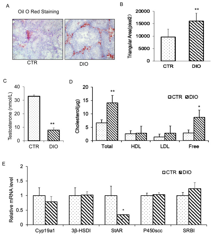
Results: It was found that there was a significant increase in body weight among DIO mice, however, the food intake showed no difference compared to control mice fed a normal diet (CTR). The number of offspring in DIO mice decreased, but there was no significant difference from the CTR group. The levels of follicle-stimulating hormone were lower in DIO mice and their luteinizing hormone levels were similar. The results showed a remarkable decrease in sperm density and motility among DIO mice. We also found that fat accumulation affected the meiosis process, mainly reflected in the cross-exchange of homologous chromosomes. In addition, overweight increased fat deposition in the testis and reduced the expression of testosterone synthesis-related enzymes, thereby affecting the synthesis and secretion of testosterone by testicular Leydig cells.

Conclusion: Fat accumulation in the testes causes testicular cell dysfunction, which affects testosterone hormone synthesis and ultimately affects sperm formation.

Keywords: Cholesterol Metabolism; Overweight; Spermatogenesis; Testosterone.


Conflict of interest statement

CONFLICT OF INTEREST

We certify that there is no conflict of interest with any financial organization regarding the material discussed in the manuscript.

Figures

Figure 1
Figure 1
The result of constructing overweight mouse. (A) Photographs of mice at age 15 weeks; they consumed either high-fat or control diet since the age of 4 weeks. (B) Intake difference in mice consuming the high fat vs control diet at age 15 weeks. (C) Body weight analysis of 20 mice for each group. (D) Comparison of the number of offspring in control and overweight mice. * p<0.05.
Figure 2
Figure 2
The estimation of sperm motility and semen density. (A) Mouse sperm motility in the control group and in the DIO group. Different colors describe the different states of motions of the sperm. (B) The comparison of progressive motility in the control and DIO mice. (C) Semen density analysis of control and DIO mice. DIO, diet-induced overweight. * p<0.05.
Figure 3
Figure 3
Histological analysis of the seminiferous tubules and caudal epididymis and immunohistochemistry (IHC) analysis of the control and overweight mice. (A) Histological analysis of the caudal epididymis of the control and overweight mice. Scale bars, 200 μm. The control and overweight mice were 15-weeks-old. (B) Relative epididymal sperm Density of the two groups. (C) Histological analysis of the seminiferous tubules of the control and overweight mice. Scale bars, 100 μm. (D) The thickness of the seminiferous epithelium of the two groups. (E) VASA IHC analysis of the germ cells in control and overweight mice depicted the number of germ cells. (F) Quantification of germ cells in the seminiferous tubules of the two groups. * p<0.05.
Figure 4
Figure 4
Observation of four periods of meiosis of spermatocytes. (A) Spermatocytes were stained for γH2AX (red) and SYCP3 (green) in the control group and DIO group. Scale bars, 10 μm. (B) Spermatocytes were stained for SYCP3 (red), SYCP1(green), and DAPI (blue) in the control group and DIO group. Scale bars, 10 μm. (C) Statistics of spermatocytes in DIO group and control group at each stage of meiosis. γH2AX, H2A histone family member X; SYCP3, synaptonemal complex protein 3; DIO, diet-induced overweight; DAPI, 4′-6-diamidino-2-phenylindole.
Figure 5
Figure 5
Analysis of expression of MLH1 during the DNA mismatch repair in sperm meiosis. (A) Control and overweight mice spermatocytes were stained for SYCP3 (red) and MLH1 (green). Scale bars, 10 μm. (B) The location of mouse spermatocyte MLH1 and the crossover site statistics. MLH1, MutL homolog 1; SYCP3, synaptonemal complex protein 3. * p<0.05.
Figure 6
Figure 6
Fat deposition in the testis and the effects of fat deposition on the synthesis of testosterone. (A) Oil red staining of frozen sections of testicular tissue in the control group and DIO group. (B) Oil red coloring area comparison in the two groups. (C) Comparison of testicular testosterone levels. (D) Different types of cholesterol levels in mouse testes. (E) Expression of key genes in testosterone synthesis. DIO, diet-induced overweight. * p<0.05, ** p<0.01.

Similar articles

Cited by

References

    1. Pini T, Raubenheimer D, Simpson SJ, Crean AJ. Obesity and male reproduction; placing the western diet in context. Front Endocrinol (Lausanne) 2021;12:622292. doi: 10.3389/fendo.2021.622292. - DOI - PMC - PubMed
    1. Schwander B, Nuijten M, Evers S, Hiligsmann M. Replication of published health economic obesity models: assessment of facilitators, hurdles and reproduction success. Pharmacoeconomics. 2021;39:433–46. doi: 10.1007/s40273-021-01008-7. - DOI - PMC - PubMed
    1. Piche ME, Tchernof A, Despres JP. Obesity phenotypes, diabetes, and cardiovascular diseases. Circ Res. 2020;126:1477–500. doi: 10.1161/CIRCRESAHA.120.316101. - DOI - PubMed
    1. Luo R, Chen L, Song X, et al. Possible role of GnIH as a novel link between hyperphagia-induced obesity-related metabolic derangements and hypogonadism in male mice. Int J Mol Sci. 2022;23:8066. doi: 10.3390/ijms23158066. - DOI - PMC - PubMed
    1. Yi X, Gao H, Chen D, et al. Effects of obesity and exercise on testicular leptin signal transduction and testosterone biosynthesis in male mice. Am J Physiol Regul Integr Comp Physiol. 2017;312:R501–10. doi: 10.1152/ajpregu.00405.2016. - DOI - PubMed

LinkOut - more resources