Quantitative analysis of zirconia and titanium implant artefacts in three-dimensional virtual models of multi-slice CT and cone beam CT: does scan protocol matter?
- PMID: 37641962
- PMCID: PMC10968770
- DOI: 10.1259/dmfr.20230275
Quantitative analysis of zirconia and titanium implant artefacts in three-dimensional virtual models of multi-slice CT and cone beam CT: does scan protocol matter?
Abstract
Objectives: Artefacts from dental implants in three-dimensional (3D) imaging may lead to incorrect representation of anatomical dimensions and impede virtual planning in navigated implantology. The aim of this study was quantitative assessment of artefacts in 3D STL models from cone beam CT (CBCT) and multislice CT (MSCT) using different scanning protocols and titanium-zirconium (Ti-Zr) and zirconium (ZrO2) implant materials.
Methods: Three ZrO2 and three Ti-Zr implants were respectively placed in the mandibles of two fresh human specimens. Before (baseline) and after implant placement, 3D digital imaging scans were performed (10 repetitions per timepoint: voxel size 0.2 mm³ and 0.3 mm³ for CBCT; 80 and 140 kV in MSCT). DICOM data were converted into 3D STL models and evaluated in computer-aided design software. After precise merging of the baseline and post-op models, the surface deviation was calculated, representing the extent of artefacts in the 3D models.
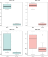
Results: Compared with baseline, ZrO2 emitted 36.5-37.3% (±0.6-0.8) artefacts in the CBCT and 39.2-50.2% (±0.5-1.2) in the MSCT models. Ti-Zr implants produced 4.1-7.1% (±0.3-3.0) artefacts in CBCT and 5.4-15.7% (±0.5-1.3) in MSCT. Significantly more artefacts were found in the MSCT vs CBCT models for both implant materials (p < 0.05). Significantly fewer artefacts were visible in the 3D models from scans with higher kilovolts in MSCT and smaller voxel size in CBCT.
Conclusions: Among the four applied protocols, the lowest artefact proportion of ZrO2 and Ti-Zr implants in STL models was observed with CBCT and the 0.3 mm³ voxel size.
Keywords: artifact; computed tomography; cone-beam computed tomography; dental implant; three-dimensional imaging; titanium; zirconium.
Figures


Similar articles
-
Assessment of peri-implant defects at titanium and zirconium dioxide implants by means of periapical radiographs and cone beam computed tomography: An in-vitro examination.Clin Oral Implants Res. 2018 Dec;29(12):1195-1201. doi: 10.1111/clr.13383. Epub 2018 Nov 22. Clin Oral Implants Res. 2018. PMID: 30387207
-
Influence of Metal Artifact Reduction Tool of Two Cone Beam CT on the Detection of Bone Graft Loss Around Titanium and Zirconium Implants-An Ex Vivo Diagnostic Accuracy Study.Clin Oral Implants Res. 2025 Mar;36(3):279-289. doi: 10.1111/clr.14381. Epub 2024 Nov 19. Clin Oral Implants Res. 2025. PMID: 39560377
-
Cone beam computed tomography artefacts around dental implants with different materials influencing the detection of peri-implant bone defects.Clin Oral Implants Res. 2020 Jul;31(7):595-606. doi: 10.1111/clr.13596. Epub 2020 Mar 23. Clin Oral Implants Res. 2020. PMID: 32147872
-
Magnetic resonance imaging in zirconia-based dental implantology.Clin Oral Implants Res. 2015 Oct;26(10):1195-202. doi: 10.1111/clr.12430. Epub 2014 Jun 4. Clin Oral Implants Res. 2015. PMID: 24893967
-
Cone beam computed tomography in implant dentistry: recommendations for clinical use.BMC Oral Health. 2018 May 15;18(1):88. doi: 10.1186/s12903-018-0523-5. BMC Oral Health. 2018. PMID: 29764458 Free PMC article. Review.
Cited by
-
Evaluation of the effect of reducing metal artifacts in multi-detector CT imaging of zirconia and titanium implants.Oral Radiol. 2025 Jul;41(3):421-429. doi: 10.1007/s11282-025-00814-5. Epub 2025 Mar 4. Oral Radiol. 2025. PMID: 40038165
References
MeSH terms
Substances
LinkOut - more resources
Full Text Sources
Miscellaneous

