

Does cone beam CT change the treatment decision for maxillary second and third molars? A prospective study
- PMID: 37641963
- PMCID: PMC10552123
- DOI: 10.1259/dmfr.20230128
Does cone beam CT change the treatment decision for maxillary second and third molars? A prospective study
Abstract
Objectives: To evaluate whether information from CBCT changes the treatment plan for maxillary second and third molars and to examine clinical and radiographic parameters with an impact on treatment decision.
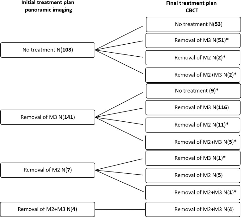
Methods: This prospective study included 260 maxillary third molars with superimposition onto the second molar in panoramic images (170 patients; mean age 28 years, range 16-63). An initial treatment plan was based on clinical findings and panoramic images. After CBCT, a final treatment plan was decided. Treatment was undertaken based on the final treatment plan. Through logistic regression analyses, impact of clinical and radiographic parameters on change in treatment plan, removal of the third molar vs no treatment, and removal of the second vs third molar were evaluated.
Results: The treatment plan changed in 82 cases (32%). Sixteen cases (6%) changed from removal of the third molar to removal of the second molar. Regression analyses showed that severe resorption in the second molar was significantly related to a change in treatment plan. Removal of a third molar was decided in 180 cases and regression analyses identified that mesioangulation of the third molar, marginal bone loss, superficial resorption, and age were significantly related to removal of the third molar vs no treatment. Thirty second molars were removed, and regression analyses showed that severe resorption was significantly related to removal of the second molar instead of the third molar.
Conclusions: Parameters such as resorption evaluated in CBCT can modify the treatment decision, resulting in removal of the second and/or the third molar.
Keywords: Cone Beam Computed Tomography; Diagnostic Imaging; Maxilla; Panoramic Radiography; Third Molar.
Figures
References
-
- European Commision . European guidelines on radiation protection in dental radiology: The safe use of radiographs in dental practice . In: Radiation protection. European Commision; 2004.
-
- Moreira-Souza L, Butini Oliveira L, Gaêta-Araujo H, Almeida-Marques M, Asprino L, Oenning AC. Comparison of Cbct and panoramic radiography for the assessment of bone loss and root Resorption on the second molar associated with third molar Impaction: A systematic review. Dentomaxillofac Radiol 2022; 51(): 20210217. doi: 10.1259/dmfr.20210217 - DOI - PMC - PubMed
MeSH terms
LinkOut - more resources
Full Text Sources
