. 2023 Sep 5;12(17):e028185.
doi: 10.1161/JAHA.122.028185. Epub 2023 Aug 29.

TRAF Family Member 4 Promotes Cardiac Hypertrophy Through the Activation of the AKT Pathway

Affiliations

TRAF Family Member 4 Promotes Cardiac Hypertrophy Through the Activation of the AKT Pathway

Jian Li et al. J Am Heart Assoc. .

Abstract

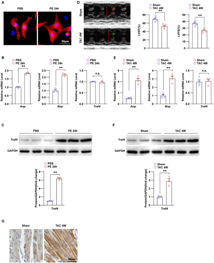
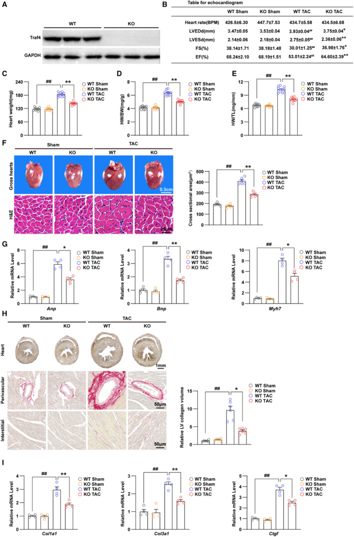
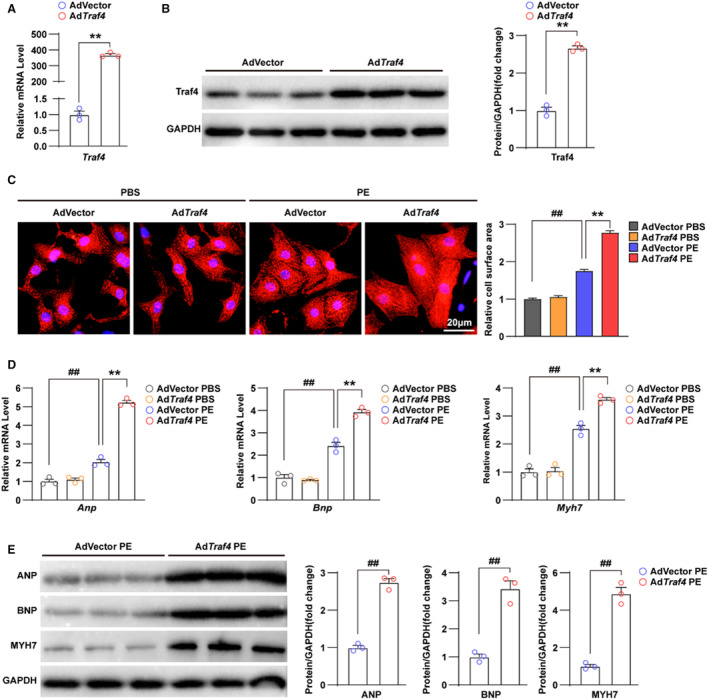
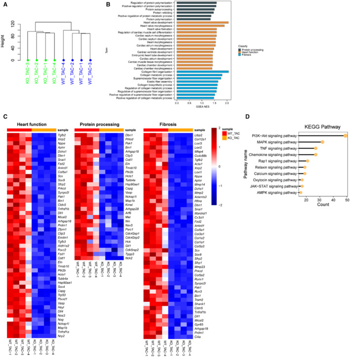
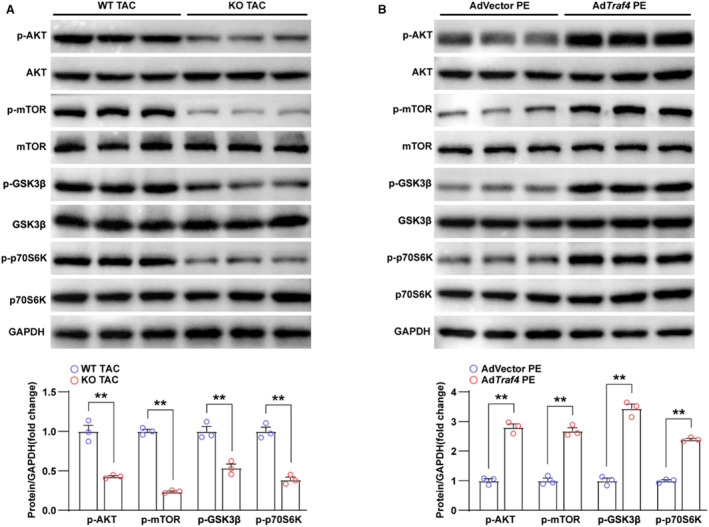
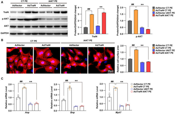
Background Pathological cardiac hypertrophy is a major cause of heart failure morbidity. The complex mechanism of intermolecular interactions underlying the pathogenesis of cardiac hypertrophy has led to a lack of development and application of therapeutic methods. Methods and Results Our study provides the first evidence that TRAF4, a member of the tumor necrosis factor receptor-associated factor (TRAF) family, acts as a promoter of cardiac hypertrophy. Here, Western blotting assays demonstrated that TRAF4 is upregulated in cardiac hypertrophy. Additionally, TRAF4 deletion inhibits the development of cardiac hypertrophy in a mouse model after transverse aortic constriction surgery, whereas its overexpression promotes phenylephrine stimulation-induced cardiomyocyte hypertrophy in primary neonatal rat cardiomyocytes. Mechanistically, RNA-seq analysis revealed that TRAF4 promoted the activation of the protein kinase B pathway during cardiac hypertrophy. Moreover, we found that inhibition of protein kinase B phosphorylation rescued the aggravated cardiomyocyte hypertrophic phenotypes caused by TRAF4 overexpression in phenylephrine-treated neonatal rat cardiomyocytes, suggesting that TRAF4 may regulate cardiac hypertrophy in a protein kinase B-dependent manner. Conclusions Our results revealed the regulatory function of TRAF4 in cardiac hypertrophy, which may provide new insights into developing therapeutic and preventive targets for this disease.

Keywords: AKT pathway; TRAF4; cardiac hypertrophy; heart failure; phenylephrine; primary neonatal rat cardiomyocytes; transverse aortic constriction.


Figures

Figure 1
Figure 1. TRAF4 expression is upregulated in cardiac hypertrophy models.
A, Representative immunofluorescence images of α‐actinin (red)‐ and DAPI (blue)‐stained NRCMs treated with phosphate buffered saline (PBS) or phenylephrine (PE) for 24 h, respectively. B, qPCR analysis of the mRNA levels of hypertrophic markers (atrial natriuretic peptide [Anp] and brain natriuretic peptide [Bnp]) and tumor necrosis factor receptor–associated factor 4 (Traf4) in NRCMs treated with PBS or PE for 24 h. C, WB analysis for protein levels of TRAF4 in NRCMs treated with PBS or PE for 24 h. D, Representative echocardiographic images and left ventricular fraction shortening (LVFS) and left ventricular ejection fraction (LVEF) statistics of mice at 4 weeks after sham or transverse aortic constriction (TAC) surgery (n=5 mice in each group). E, qPCR analysis of the RNA levels of hypertrophic markers and Traf4 in left ventricular tissues of mice at 4 weeks after sham or TAC surgery (n=4 mice in each group). F, WB analysis for protein levels of TRAF4 in the myocardium of mice at 4 weeks after sham or TAC surgery (n=3 mice in each group), and GAPDH served as a loading control. G, Immunohistochemistry images of TRAF4 in mice heart tissues of the sham and TAC group (n=4 mice in each group). Statistical analysis was carried out with 2‐tailed Student t test. **P<0.01 vs PBS or sham group. DAPI indicates 4,6‐diamidino‐2‐phenylindole; NRCMs, neonatal rat cardiomyocytes; n.s., not significant; qPCR, quantitative polymerase chain reaction; TAC, transverse aortic constriction; and WB, Western blotting.
Figure 2
Figure 2. TRAF4 deficiency ameliorates TAC‐induced cardiac hypertrophy.
A, WB analysis for protein levels of TRAF4 (tumor necrosis factor receptor–associated factor 4) in myocardium tissues from wild‐type (WT) and Traf4‐knock‐out (KO) mice (n=3 mice/group), and GAPDH served as a loading control. B, Echocardiogram parameters (left ventricular end‐diastolic dimension [LVEDd], left ventricular end‐systolic dimension [LVESd], fraction shortening [FS] ejection fraction [EF]) of WT and Traf4‐KO mice 4 weeks after sham or transverse aortic constriction (TAC) surgery (n=10 mice/group). Heart weight (C), heart weight (HW)/body weight (BW) ratio (D), and HW/tibia length (TL) ratio (E) in WT and Traf4‐KO mice 4 weeks after sham or TAC surgery (n=10 mice/group). F, Representative images of gross hearts, hematoxylin and eosin (H&E)–stained sections from the left ventricle (LV) (left) and quantitative cross‐sectional area based on H&E staining (right) of WT and Traf4‐KO mice 4 weeks after sham or TAC surgery (n=6 mice/group). G, The mRNA levels of hypertrophic markers (atrial natriuretic peptide [Anp], brain natriuretic peptide [Bnp], and myosin heavy chain 7 [Myh7]) in myocardium of WT and Traf4‐KO mice 4 weeks after sham or TAC surgery (n=4 mice/group). H, Representative images of perivascular and interstitial PSR‐stained LV sections (left) and quantitative LV collagen volume base on PSR staining (right) of WT and Traf4‐KO mice at 4 weeks after sham or TAC surgery (n=6 mice/group). I, qPCR analysis of mRNA levels of collagen synthesis–related genes (collagen type I alpha 1(Col1a1), collagen type III alpha 1(Col3a1), and connective tissue growth factor (Ctgf)) in myocardium tissues from WT and Traf4‐KO mice at 4 weeks after sham or TAC surgery (n=4 mice/group). Statistical analysis was carried out by 2‐way ANOVA. ## P<0.01 vs WT sham; *P<0.05 or **P<0.01 vs WT TAC. PSR indicates picrosirius red; qPCR, quantitative polymerase chain reaction; and WB, Western blotting.
Figure 3
Figure 3. Overexpression of TRAF4 exacerbates phenylephrine‐induced cardiomyocyte hypertrophy.
A, qPCR analysis of mRNA level of Traf4 (tumor necrosis factor receptor–associated factor 4) in adenovirus vector (AdVector) and AdTraf4 infected NRCMs. B, WB analysis for protein level of TRAF4 in AdVector and AdTraf4 infected NRCMs, and GAPDH served as a loading control. C, Representative immunofluorescence images (left) and quantitative relative cell surface area (right) of α‐actinin (red)‐ and DAPI (blue)–stained AdVector and AdTraf4 infected NRCMs treated with phosphate buffered saline (PBS) or phenylephrine (PE) for 24 h. D, qPCR analysis for mRNA levels and (E) WB analysis for protein levels of hypertrophic markers (Atrial natriuretic peptide (Anp), Brain natriuretic peptide (Bnp) and Myosin heavy chain 7(Myh7)) in AdVector and AdTraf4 infected NRCMs treated with PBS or PE for 24 h, respectively. Statistical analysis of (A, B, and E) was carried out by 2‐tailed Student t test, and (C and D) was carried out by nonparametric Kruskal–Wallis test and 2‐way ANOVA, respectively. ## P<0.01 vs AdVector PBS and **P<0.01 vs AdVector PE. DAPI indicates 4,6‐diamidino‐2‐phenylindole; NRCMs, neonatal rat cardiomyocytes; qPCR, quantitative polymerase chain reaction; TRAF4, tumor necrosis factor receptor–associated factor 4; and WB, Western blotting.
Figure 4
Figure 4. RNA‐seq analysis reveals that TRAF4 influences AKT‐related pathways.
A, Clustering analysis result of the RNA‐seq data from wild‐type (WT) and tumor necrosis factor receptor–associated factor 4 (Traf4)‐knock‐out (KO) mice left ventricular tissues 4 weeks after transverse aortic constriction (TAC) surgery. B, Gene Set Enrichment Analysis (GSEA) pathway enrichment analysis of normalized enrichment score (NES) in pathways related to myocardial function, protein processing, and fibrosis. C, Heatmaps of downregulated myocardial function–related, protein processing–related, and fibrosis gene expression profiles based on the RNA‐seq data set. D, Kyoto Encyclopedia of Genes and Genomes (KEGG) pathway enrichment analysis based on the RNA‐seq data set (PI3K‐AKT [phosphatidylinositol 3‐kinase‐protein kinase B], MAPK [mitogen activated kinase‐like protein], TNF [tumor necrosis factor], JAK–STAT [Janus kinase‐signal transducer and activator of transcription], AMPK [AMP‐activated protein kinase]). n=4 mice in each group. RNA‐seq indicates RNA sequencing.
Figure 5
Figure 5. TRAF4 overexpression promotes the activation of AKT signaling pathway.
A, WB analysis of protein levels of total and p‐AKT (phosphorylated protein kinase B) pathway‐related proteins (p‐AKT, p‐GSK3β [phosphorylated glycogen synthase kinase 3 beta], p‐mTOR [phosphorylated mammalian target of rapamycin], p‐P70S6K [phosphorylated ribosomal protein S6 kinase], AKT, GSK3β, mTOR, and P70S6K) in left ventricular tissues from wild‐type (WT) and tumor necrosis factor receptor–associated factor 4 (Traf4) knock‐out (KO) mice at 4 weeks after transverse aortic constriction (TAC) surgery; GAPDH served as a loading control. B, Protein levels of total and phosphorylated AKT pathway‐related proteins in adenovirus vector (AdVector)‐ and AdTraf4‐infected NRCMs treated with phenylephrine (PE) for 24 h. Statistical analysis was carried out by 2‐tailed Student t test. **P<0.01 vs WT TAC or AdVector PE. NRCMs indicates neonatal rat cardiomyocytes; TRAF4, tumor necrosis factor receptor–associated factor 4; and WB, Western blotting.
Figure 6
Figure 6. AKT inhibitor abrogates the promoting effect of TRAF4 overexpression on cardiac hypertrophy.
A, Protein levels of Traf4 (tumor necrosis factor receptor–associated factor 4), total AKT (protein kinase B), and phosphorylated AKT (p‐AKT) in DMSO‐treated control (CT) or inhibitors of AKT phosphorylation (iAKT)‐treated adenovirus vector (AdVector)‐ and AdTraf4‐infected NRCMs treated with phenylephrine (PE) for 24 h; GAPDH served as a loading control. B, Representative immunofluorescence images of α‐actinin (red)‐ and DAPI (blue)–stained CT or iAKT‐treated AdVector‐ and AdTraf4‐infected NRCMs treated with PE for 24 h. C, mRNA levels of hypertrophic markers (atrial natriuretic peptide (Anp), brain natriuretic peptide (Bnp), and myosin heavy chain 7(Myh7)) in DMSO‐ or iAKT‐treated AdVector‐ and AdTraf4‐infected NRCMs treated with PE for 24 h. Statistical analysis was carried out by 2‐ way ANOVA. ## P<0.01 vs AdVector CT PE; **P<0.01 vs AdTraf4 CT PE. DAPI indicates 4,6‐diamidino‐2‐phenylindole; DMSO, dimethyl sulfoxide; and NRCMs, neonatal rat cardiomyocytes.

Similar articles

Cited by

References

    1. Rame JE, Dries DL. Heart failure and cardiac hypertrophy. Curr Treat Options Cardiovasc Med. 2007;9:289–301. doi: 10.1007/s11936-007-0024-3 - DOI - PubMed
    1. Nakamura M, Sadoshima J. Mechanisms of physiological and pathological cardiac hypertrophy. Nat Rev Cardiol. 2018;15:387–407. doi: 10.1038/s41569-018-0007-y - DOI - PubMed
    1. Miao R, Lu Y, Xing X, Li Y, Huang Z, Zhong H, Huang Y, Chen AF, Tang X, Li H, et al. Regulator of G‐protein signaling 10 negatively regulates cardiac remodeling by blocking mitogen‐activated protein kinase‐extracellular signal‐regulated protein kinase 1/2 signaling. Hypertension. 2016;67:86–98. doi: 10.1161/HYPERTENSIONAHA.115.05957 - DOI - PubMed
    1. Savarese G, Lund LH. Global public health burden of heart failure. Card Fail Rev. 2017;3:7–11. doi: 10.15420/cfr.2016:25:2 - DOI - PMC - PubMed
    1. Frey N, Katus HA, Olson EN, Hill JA. Hypertrophy of the heart: a new therapeutic target? Circulation. 2004;109:1580–1589. doi: 10.1161/01.CIR.0000120390.68287.BB - DOI - PubMed

Publication types

Substances