. 2023 Oct;124(10):1530-1545.
doi: 10.1002/jcb.30462. Epub 2023 Aug 29.

Electrical stimulation alters DNA methylation and promotes neurite outgrowth

Affiliations

Electrical stimulation alters DNA methylation and promotes neurite outgrowth

Ajay Ashok et al. J Cell Biochem. 2023 Oct.

Abstract

Electrical stimulation (ES) influences neural regeneration and functionality. We here investigate whether ES regulates DNA demethylation, a critical epigenetic event known to influence nerve regeneration. Retinal ganglion cells (RGCs) have long served as a standard model for central nervous system neurons, whose growth and disease development are reportedly affected by DNA methylation. The current study focuses on the ability of ES to rescue RGCs and preserve vision by modulating DNA demethylation. To evaluate DNA demethylation pattern during development, RGCs from mice at different stages of development, were analyzed using qPCR for ten-eleven translocation (TETs) and immunostained for 5 hydroxymethylcytosine (5hmc) and 5 methylcytosine (5mc). To understand the effect of ES on neurite outgrowth and DNA demethylation, cells were subjected to ES at 75 µAmp biphasic ramp for 20 min and cultured for 5 days. ES increased TETs mediated neurite outgrowth, DNA demethylation, TET1 and growth associated protein 43 levels significantly. Immunostaining of PC12 cells following ES for histone 3 lysine 9 trimethylation showed cells attained an antiheterochromatin configuration. Cultured mouse and human retinal explants stained with β-III tubulin exhibited increased neurite growth following ES. Finally, mice subjected to optic nerve crush injury followed by ES exhibited improved RGCs function and phenotype as validated using electroretinogram and immunohistochemistry. Our results point to a possible therapeutic regulation of DNA demethylation by ES in neurons.

Keywords: H3K9Me3; TET1; electrical stimulation; epigenetics; nerve regeneration; retinal ganglion cells.


Conflict of interest statement

CONFLICT OF INTEREST STATEMENT

Dong Chen is scientific founder of FireCyte Therapeutics and a consultant of i-Lumen Scientific and Sichuan PriMed. Kin-Sang Cho is a consultant in FireCyte Therapeutics. Ajay Ashok, Anton Lennikov, Karen Chang, Dong Chen, Kin-Sang Cho, Wai Lydia Tai, and Tor Paas Utheim are inventors of a patent application for using microcurrent electrical stimulation technology or nerve regenerative approach to treat eye diseases.

Figures

FIGURE 1
FIGURE 1
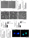
Nerve growth factor induces PC12 differentiation and TET1 expression. (A) Bright field images of PC-12 cells exposed to increasing concentrations of NGF (0, 5, and 100 ng/mL) exhibited a dose dependent increase in differentiation as indicated by significantly increased neurite outgrowth quantified by (B) neurite length and (C) neurite numbers per field. Images were acquired 5 days post NGF induction of differentiation. (D) Results of qPCR assessments of the levels of TET1, TET2, and TET3 in PC-12 cells exposed to increasing concentrations of NGF. A significant dose dependent increase in TET1 mRNA was observed in PC12 cells. RNA samples were collected 24 h post NGF treatment for qPCR analysis. Values are means ± SEM of indicated number of samples (n). *p ≤ 0.05; ***p ≤ 0.001. mRNA, messenger RNA; NGF, nerve growth factor.
FIGURE 2
FIGURE 2
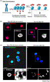
Electrical stimulation increased neurite outgrowth and DNA demethylation in PC-12 cells. (A–C) ES increased neurite outgrowth in PC12 cells compared to control unstimulated cells. PC12 cells incubated with a low level of NGF (5 ng/mL) (panels 1 and 2) and received 20 min ES at 24 h post NGF induction. (D–F) ES increased neurite outgrowth in PC12 cells compared to control unstimulated cells. PC12 cells incubated with a low level of NGF (5 ng/mL) (panels 1 and 2) and received 20 min ES at 24 h post NGF induction. However, pre-incubating PC12 cells with TETi76 diethyl ester (a pan TET inhibitor) at 12.5 μM (for 16 h) before ES (panel 3), blocked the ES induced neurite outgrowth. Result of qPCR assessing mRNA levels of GAP43 (G) and TET1 (H) expression in Non-ES- and ES-treated cells. (I) Immunocytochemistry based analysis of 5hmc (green) expression, an intermediate product of DNA demethylation process, reveals increased expression of the substrate following ES. The expression was localized in the nucleus (blue). Values are means ± SEM. **p ≤ 0.01; *p ≤ 0.05 by the Student t test. mRNA, messenger RNA; NGF, nerve growth factor.
FIGURE 3
FIGURE 3
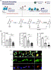
Electrical stimulation reduced heterochromatin configuration formation. (A) Graphical illustration (Biorender was used to create the illustration) showing the histone distancing following increased TET1 and DNA demethylation, thereby promoting translational activity. Immunolabeling of H3K9me3 (green) used as a marker to reveal the distribution of histone-DNA complex in the nuclei. Note PC12 cells treated with ES after (B) 20 min (panels 1 and 2) did not show any significant changes in H3K9me3 localization. However, PC12 cells treated with ES after (C) 24 h culturing (panels 1 and 3) showed periphery and distanced localized H3K9me3 with a distinctive (panels 3 and 4) translational pattern compared to non-ES-treated cells. ES condition: 75 μAmp-biphasic/ramp/50 ms for 20 min-once. ES, electrical stimulation.
FIGURE 4
FIGURE 4
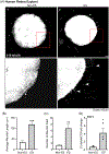
Developmental downregulation of TETs in RGCs. (A) Graphical representation of DNA demethylation mechanism and its effect on chromatin structure and translational activity. *thymine-DNA glycosylase (TDG)-mediated base excision repair (BER) (Biorender was used to create the illustration). (B) Quantification of TET1, TET2, and TET3 mRNA with qPCR in the developing mouse RGCs. Note the significantly decreased mRNA levels of all TET(s) with age. Retinas from 4 mice were pooled into two groups (represented as two data points) for the E-14 age group. (C) Images of retinal sections of E-16, P-0 and adult mice (3 months) double-immunolabeled with primary antibodies against 5mc (red) and 5hmc (green); retinal sections were counterstained with a nuclear marker DAPI (blue). Values are means ± SEM. *p ≤ 0.05; **p ≤ 0.01; ***p ≤ 0.001 by the Student t test. GCL, ganglion cell layer; INL, inner nuclear layer; mRNA, messenger RNA.
FIGURE 5
FIGURE 5
Electrical stimulation increased neurite outgrowth in mouse retinal explant cultures. (A) Immunodetection of β-III tubulin-labeled neurites (arrowhead) in representative adult mouse retinal explant cultures with and without ES. (B, C) Quantification using LASX software reveals significantly longer (B) and increased number of neurites in cultures subjected to ES compared to unstimulated cultures. After 24 h in culture in trans-well membranes, the tissues were subjected to ES. The images were presented in grayscale (panels 1 and 2) for optimal and clear presentation and analysis. Values are mean ± SEM. *p < 0.05 by the Student t test. ES, electrical stimulation.
FIGURE 6
FIGURE 6
Electrical stimulation increased neurite outgrowth in human retinal explant cultures. (A) Immunodetection of β-III tubulin (to label neurites) in adult human retinal explant cultures followed by quantification of neurite outgrowth using LASX software. Significantly (B) longer (arrowheads) and (C) a greater number of neurites in retinal explant cultures exposed to ES than in unstimulated cultures was noted. Human and mouse retinal explants underwent identical culture and experimental conditions. (D) Results of qPCR quantification of TET1 exhibited a significant increase in TET1 mRNA in the retinal explants undergone ES versus non-ES controls. The images were presented in grayscale (panels 1 and 2) for optimal and clear presentation and analysis. Values are mean ± SEM of the indicated n. *p < 0.05; **p < 0.01; ***p < 0.001 by the Student t test. ES, electrical stimulation; mRNA, messenger RNA.
FIGURE 7
FIGURE 7
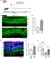
Electrical stimulation alleviates RGC damage and functional deterioration in ONC model. (A) Graphical abstract of the in vivo experimental plan (Biorender was used to create the illustration). (B) Representative images of immunohistochemistry of Thy1-YFP mouse retinal sections showing YFP+ RGCs (green; cells in the innermost layer) bearing increased nerve fibers in the ES group (arrowheads). (C) Quantification of YFP+ RGCs exhibiting axonal growth in ES and non-ES mice group retinal sections. ***p < 0.001 by Student t test. (D–E) Retinal sections from non-ES and ES-treated Thy1-YFP transgenic mice immunostained for 5hmc. Number of cells co-expressing Thy1-YFP and 5hmc in the ES mice group were higher, but much less seen in non-ES mouse retinas. (F) ES improves ERG responses in ONC mice. Following ONC, the pSTR values dropped significantly compared to noninjured control eye (Cnt); ES-treatment improved the ERG pSTR amplitude. Values are mean ± SEM. *p < 0.05; **p < 0.01 by the Student t test. ES, electrical stimulation; GCL, ganglion cell layer; INL, inner nuclear layer; ONL, outer nuclear layer; IPL, inner plexiform layer; RGC, retinal ganglion cells.

References

    1. Quigley HA. Understanding glaucomatous optic neuropathy: the synergy between clinical observation and investigation. Annu Rev Vis Sci. 2016;2:235–254. - PubMed
    1. Williams PR, Benowitz LI, Goldberg JL, He Z. Axon regeneration in the mammalian optic nerve. Annu Rev Vis Sci. 2020;6:195–213. - PubMed
    1. Ashok A, Pooranawattanakul S, Tai WL, et al. Epigenetic regulation of optic nerve development, protection, and repair. Int J Mol Sci. 2022;23:8927. - PMC - PubMed
    1. Lu Y, Brommer B, Tian X, et al. Reprogramming to recover youthful epigenetic information and restore vision. Nature. 2020;588:124–129. - PMC - PubMed
    1. Moyon S, Frawley R, Marechal D, et al. TET1-mediated DNA hydroxymethylation regulates adult remyelination in mice. Nat Commun. 2021;12:3359. - PMC - PubMed

LinkOut - more resources