. 2023 Aug 29;11(5):e0083323.
doi: 10.1128/spectrum.00833-23. Online ahead of print.

Emergence of the Dickeya genus involved duplication of the OmpF porin and the adaptation of the EnvZ-OmpR signaling network

Affiliations

Emergence of the Dickeya genus involved duplication of the OmpF porin and the adaptation of the EnvZ-OmpR signaling network

Clémence Cochard et al. Microbiol Spectr. .

Abstract

Genome evolution, and more specifically gene duplication, is a key process shaping host-microorganism interaction. The conserved paralogs usually provide an advantage to the bacterium to thrive. If not, these genes become pseudogenes and disappear. Here, we show that during the emergence of the genus Dickeya, the gene encoding the porin OmpF was duplicated. Our results show that the ompF2 expression is deleterious to the virulence of Dickeya dadantii, the agent causing soft rot disease. Interestingly, ompF2 is regulated while ompF is constitutive but activated by the EnvZ-OmpR two-component system. In vitro, acidic pH triggers the system. The pH measured in four eudicotyledons increased from an initial pH of 5.5 to 7 within 8 h post-infection. Then, the pH decreased to 5.5 at 10 h post-infection and until full maceration of the plant tissue. Yet, the production of phenolic acids by the plant's defenses prevents the activation of the EnvZ-OmpR system to avoid the ompF2 expression even though environmental conditions should trigger this system. We highlight that gene duplication in a pathogen is not automatically an advantage for the infectious process and that, there was a need for our model organism to adapt its genetic regulatory networks to conserve these duplicated genes. IMPORTANCE Dickeya species cause various diseases in a wide range of crops and ornamental plants. Understanding the molecular program that allows the bacterium to colonize the plant is key to developing new pest control methods. Unlike other enterobacterial pathogens, Dickeya dadantii, the causal agent of soft rot disease, does not require the EnvZ-OmpR system for virulence. Here, we showed that during the emergence of the genus Dickeya, the gene encoding the porin OmpF was duplicated and that the expression of ompF2 was deleterious for virulence. We revealed that while the EnvZ-OmpR system was activated in vitro by acidic pH and even though the pH was acidic when the plant is colonized, this system was repressed by phenolic acid (generated by the plant's defenses). These results provide a unique- biologically relevant-perspective on the consequence of gene duplication and the adaptive nature of regulatory networks to retain the duplicated gene.

Keywords: Dickeya dadantii; EnvZ-OmpR; phenolic acids; plant pathogen; porin; two-component system.


Conflict of interest statement

The authors declare no conflict of interest.

Figures

Fig 1
Fig 1
The EnvZ-OmpR system is dispensable for virulence. (A) Representative time course of symptom development for the wild-type, ompR, envZ strains inoculated into chicory leaves. Virulence was monitored for 3 d. Extension of macerated area (B) and bacterial load (colony forming unit) (C) for the wild-type, ompR, and envZ strains inoculated into chicory leaves (n = 6). Experiments were repeated three times. (D) Representative immunoblot of the separation of OmpR and OmpR~P by SDS-PAGE Phos-Tag gel after extraction of bacteria during the infectious cycle and quantification of OmpR and OmpR~P. Cell lysate of wild type before infection and after 24, 48, and 72 h post-infection, envZ and ompR mutants from exponential growth phase culture of D. dadantii were loaded into SDS-PAGE Phos-Tag gel. Both forms of OmpR were revealed by Western blot (n = 3 biological replicates).
Fig 2
Fig 2
The pH transiently increases at the early stage of the infection and triggered the EnvZ-OmpR system. Tissue osmolarity and pH of chicory leaves (A, E, I), potatoes tuber (B and F), green pepper (C and G), and tomatoes (D and H) were measured with a nanoprobe pH sensor for pH and by vapor pressure osmometry for osmolarity (see Methods). Experiments were repeated three times. Groups with the same letter are not detectably different (P < 0.001, one-way ANOVA). (J) Representative immunoblot of the separation of OmpR and OmpR~P by SDS-PAGE Phos-Tag gel after extraction bacteria from exponential growth phase culture at different pH and quantification of OmpR and OmpR~P. Both forms of OmpR were revealed by Western blot (n = 3 biological replicates).
Fig 3
Fig 3
Expression of ompF requires EnvZ-dependent phosphorylation of OmpR. (A) Expression of ompF::uidA gene fusions at different pH in different genetic backgrounds. Bacteria were grown to the mid-log phase and lysed by sonication. The β-glucuronidase activity was measured with PNPU as a substrate. Specific activity was expressed as the change in OD405 per minute and per milligram of protein. The results are the average of three independent experiments. (B) Representative SDS-PAGE analysis of the outer membrane proteins showing the expression of the porin OmpF. A gel system supplied with 4 M urea was used and stained by Coomassie staining.
Fig 4
Fig 4
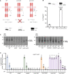
Dickeya sp. genomes contain three successive proteins. (A) Organization of the pncB-asnS-ompFs-aspC locus in Escherichia coli and in phytopathogenic bacteria from Pectobacteriaceae and Erwiniaceae family. In blue, the ompF gene, in pink and green, the ompF duplication. Rooted phylogenetic tree based on the maximum likelihood. The tree was constructed with the 16S nucleic acid sequences. (B) Schematic representation of the ompFs promoter region and in silico location of the OmpR binding sites (C) EMSA to test the binding of purified OmpR to the ompF, ompF2, and ompF3 promoter regions. As a control, DNA fragments encompassing a part of the CDS sequence of opgC were additionally present.
Fig 5
Fig 5
Expression of ompF2 is pH dependent and EnvZ-OmpR dependent and repressed in planta. (A) Schematic of the different envZ mutants and consequence on the amount of OmpR~P. (B) Representative immunoblot of the separation of OmpR and OmpR~P by SDS-PAGE Phos-Tag gel after extraction bacteria from exponential growth phase culture and quantification of OmpR and OmpR~P. Both forms of OmpR were revealed by Western blot (n = 3 biological replicates). (C) Representative SDS-PAGE analysis of the outer membrane proteins showing the expression of the porins OmpFs. A gel system supplied with 4 M urea was used and stained by Coomassie staining. (D) Expression of ompF2::uidA gene fusions at different pH in different genetic backgrounds. Bacteria were grown to the mid-log phase and lysed by sonication. B-glucuronidase activity was measured with PNPU as a substrate. Specific activity was expressed as the change in OD405 per minute and per milligram of protein. The results are the average of three independent experiments. Groups with the same letter are not detectably different (P < 0.001, one-way ANOVA).
Fig 6
Fig 6
Expression of ompF2 is repressed in planta. Expression of (A) ompF::uidA and (B) ompF2::uidA gene fusions in M63 medium and during the infection. Bacteria were lysed by sonication. The β-glucuronidase activity was measured with PNPU as a substrate. Specific activity was expressed as the change in OD405 per minute and per milligram of protein. (C) Representative SDS-PAGE analysis of the outer membrane proteins showing the expression of the porin OmpFs. A gel system supplied with 4 M urea was used and stained by Coomassie staining.
Fig 7
Fig 7
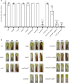
Expression of ompF2 is deleterious for D. dadantii infection. (A) Percentage of successfully infected leaves for each strain after 72 h of infection and (B) representative time course of symptom development for the wild-type, ompR, envZ, ompF, ompF2, ompF ompF2, envZ-241, envZ-241c, envZ-241 ompF2, envZ-241 ompF2c, and envZ-241 ompF strains inoculated into chicory leaves. Virulence was monitored for 3 d.
Fig 8
Fig 8
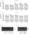
The plant phenolic acids inhibit ompF2 expression at low pH. The plant phenolic acids inhibit the EnvZ kinase activity at low pH. Expression of ompF2::uidA (A), ompF::uidA (B) gene fusions at different concentrations of salicylic acid, cinnamic acid, jasmonic acid, o-coumaric acid, and p-coumaric acid. Bacteria were grown to the mid-log phase and lysed by sonication. The β-glucuronidase activity was measured with PNPU as a substrate. Specific activity was expressed as the change in OD405 per minute and per milligram of protein. The results are the average of three independent experiments.
Fig 9
Fig 9
The reduction in ompF2 expression by the phenolic acid is due to the inhibition of the EnvZ kinase activity. (A) and (B) Representative SDS-PAGE analysis of the outer membrane proteins showing the expression of the porins OmpFs. A gel system supplied with 4 M urea was used and stained by Coomassie staining. (C) and (D) Representative immunoblot of the separation of OmpR and OmpR~P by SDS-PAGE Phos-Tag gel after extraction of bacteria from exponential growth phase culture and quantification of OmpR and OmpR~ at different concentrations of cinnamic acid and salicylic acid. Both forms of OmpR were revealed by Western blot (n = 3 biological replicates).
Fig 10
Fig 10
A proposed model for EnvZ-OmpR activation and ompFs expressions during the plant infection. (A) During D. dadantii entry in the host apoplast, the bacteria will encounter an acidic pH that should promote a high-level activation of the EnvZ-OmpR system. However, the synthesis of phenolic acids by the host cells will significantly lower the activation of the system and thus the repression of ompF2. (B) During the early phase of colonization, the bacteria will be perceived by the host cells, inducing an increase in pH and phenolic acid production. Both stimuli will maintain the low activation of EnvZ-OmpR. (C) From the last stage of colonization to the symptomatic phase, the apoplastic pH will be reverted to acidic but the production of phenolic acids will be maintained. Thus, the EnvZ-OmpR activation and so ompF2 expression will be repressed throughout the infection. During this process, the ompF expression will be assured thanks to the low level of OmpR phosphorylation. Created with BioRender.com.

References

    1. Mansfield J, Genin S, Magori S, Citovsky V, Sriariyanum M, Ronald P, Dow M, Verdier V, Beer SV, Machado MA, Toth I, Salmond G, Foster GD. 2012. Top 10 plant pathogenic bacteria in molecular plant pathology. Mol Plant Pathol 13:614–629. doi:10.1111/j.1364-3703.2012.00804.x - DOI - PMC - PubMed
    1. Reverchon S, Nasser W. 2013. Dickeya ecology, environment sensing and regulation of virulence programme. Environ Microbiol Rep 5:622–636. doi:10.1111/1758-2229.12073 - DOI - PubMed
    1. Hoch JA. 2000. Two-component and phosphorelay signal transduction. Curr Opin Microbiol 3:165–170. doi:10.1016/s1369-5274(00)00070-9 - DOI - PubMed
    1. Mizuno T, Mizushima S. 1990. Signal transduction and gene regulation through the phosphorylation of two regulatory components: the molecular basis for the osmotic regulation of the porin genes. Mol Microbiol 4:1077–1082. doi:10.1111/j.1365-2958.1990.tb00681.x - DOI - PubMed
    1. Norioka S, Ramakrishnan G, Ikenaka K, Inouye M. 1986. Interaction of a transcriptional activator, OmpR, with reciprocally osmoregulated genes, ompF and ompC, of Escherichia coli. J Biol Chem 261:17113–17119. doi:10.1016/S0021-9258(19)76006-2 - DOI - PubMed

LinkOut - more resources