

A Method for Real-Time Assessment of Mitochondrial Respiration Using Murine Corneal Biopsy
- PMID: 37642632
- PMCID: PMC10476441
- DOI: 10.1167/iovs.64.11.33
A Method for Real-Time Assessment of Mitochondrial Respiration Using Murine Corneal Biopsy
Abstract
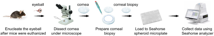
Purpose: To develop and optimize a method to monitor real-time mitochondrial function by measuring the oxygen consumption rate (OCR) in murine corneal biopsy punches with a Seahorse extracellular flux analyzer.
Methods: Murine corneal biopsies were obtained using a biopsy punch immediately after euthanasia. The corneal metabolic profile was assessed using a Seahorse XFe96 pro analyzer, and mitochondrial respiration was analyzed with specific settings.
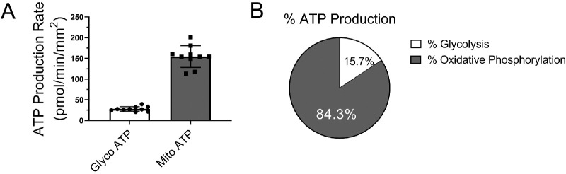
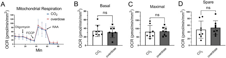
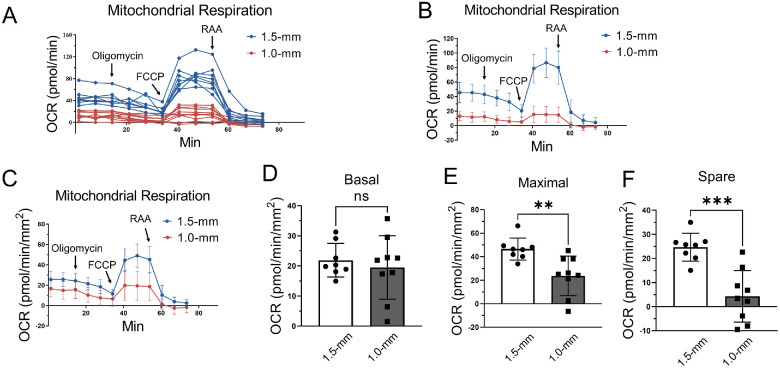
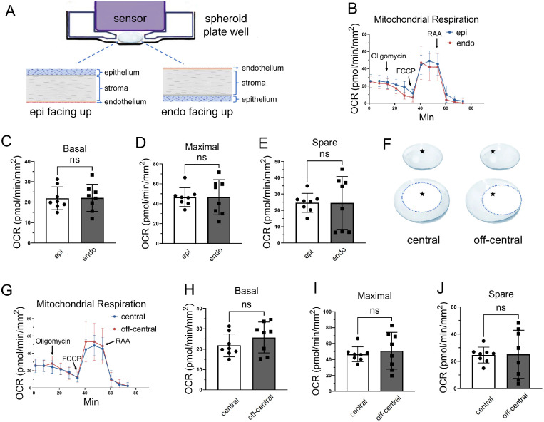
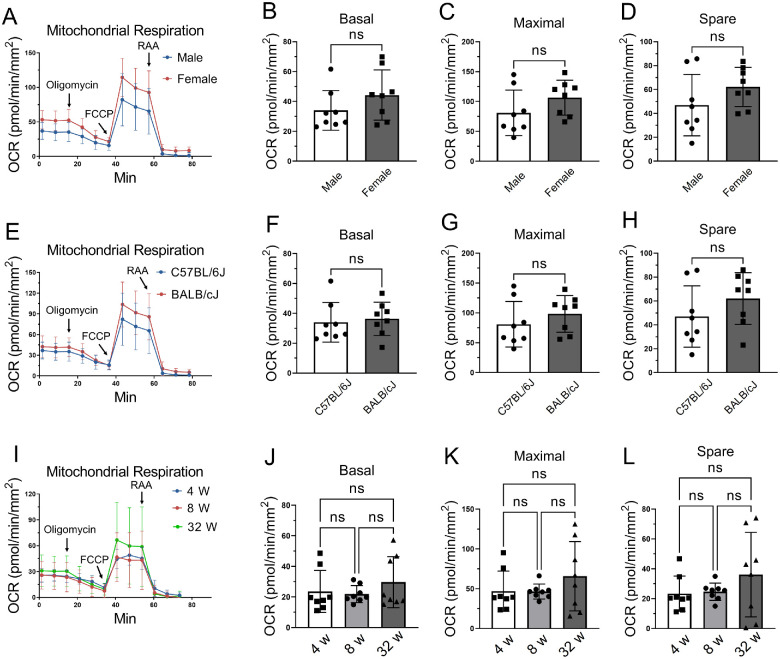
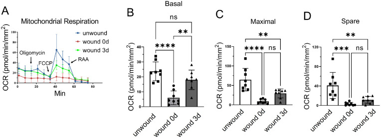
Results: Real-time adenosine triphosphate rate assay showed that mitochondrial oxidative phosphorylation is a major source of adenosine triphosphate production in ex vivo live murine corneal biopsies. Euthanasia methods (carbon dioxide asphyxiation vs. overdosing on anesthetic drugs) did not affect corneal OCR values. Mouse corneal biopsy punches in 1.5-mm diameter generated higher and more reproducible OCR values than those in 1.0-mm diameter. The biopsy punches from the central and off-central cornea did not show significant differences in OCR values. There was no difference in OCR reading by the tissue orientations (the epithelium side up vs. the endothelium side up). No significant differences were found in corneal OCR levels between sexes, strains (C57BL/6J vs. BALB/cJ), or ages (4, 8, and 32 weeks). Using this method, we showed that the wound healing process in the mouse cornea affected mitochondrial activity.
Conclusions: The present study validated a new strategy to measure real-time mitochondrial function in fresh mouse corneal tissues. This procedure should be helpful for studies of the ex vivo live corneal metabolism in response to genetic manipulations, disease conditions, or pharmacological treatments in mouse models.
Conflict of interest statement
Disclosure:
Figures
Similar articles
-
Peroxisome proliferator-activated receptor-α (PPARα) regulates wound healing and mitochondrial metabolism in the cornea.Proc Natl Acad Sci U S A. 2023 Mar 28;120(13):e2217576120. doi: 10.1073/pnas.2217576120. Epub 2023 Mar 21. Proc Natl Acad Sci U S A. 2023. PMID: 36943878 Free PMC article.
-
Monitoring Mitochondrial Respiration in Mouse Cerebellar Granule Neurons.Methods Mol Biol. 2022;2515:1-15. doi: 10.1007/978-1-0716-2409-8_1. Methods Mol Biol. 2022. PMID: 35776342
-
Real-Time Assessment of Mitochondrial Function in Cytotrophoblast and Syncytialized Trophoblast Cells Using the Seahorse XFe24 Extracellular Flux Analyzer.Methods Mol Biol. 2024;2728:137-147. doi: 10.1007/978-1-0716-3495-0_12. Methods Mol Biol. 2024. PMID: 38019398
-
Quantification of Oxygen Consumption in Retina Ex Vivo Demonstrates Limited Reserve Capacity of Photoreceptor Mitochondria.Invest Ophthalmol Vis Sci. 2015 Dec;56(13):8428-36. doi: 10.1167/iovs.15-17901. Invest Ophthalmol Vis Sci. 2015. PMID: 26747773 Free PMC article.
-
A novel high-throughput assay for respiration in isolated brain microvessels reveals impaired mitochondrial function in the aged mice.Geroscience. 2018 Aug;40(4):365-375. doi: 10.1007/s11357-018-0037-8. Epub 2018 Aug 3. Geroscience. 2018. PMID: 30074132 Free PMC article. Review.
Cited by
-
Exploring the Therapeutic Potential of Salivary Exosomes in Corneal Epithelial Wound Healing.Invest Ophthalmol Vis Sci. 2025 Aug 1;66(11):8. doi: 10.1167/iovs.66.11.8. Invest Ophthalmol Vis Sci. 2025. PMID: 40762539 Free PMC article.
-
Corneal epithelial cells upregulate macropinocytosis to engulf metabolically active axonal mitochondria released by injured axons.Ocul Surf. 2025 Jul;37:173-188. doi: 10.1016/j.jtos.2025.03.007. Epub 2025 Apr 1. Ocul Surf. 2025. PMID: 40180030
-
Mitochondria Transplantation Promotes Corneal Epithelial Wound Healing.Invest Ophthalmol Vis Sci. 2024 Jun 3;65(6):14. doi: 10.1167/iovs.65.6.14. Invest Ophthalmol Vis Sci. 2024. PMID: 38848077 Free PMC article.
References
Publication types
MeSH terms
Substances
Grants and funding
LinkOut - more resources
Full Text Sources
