. 2023 Sep 15;136(18):jcs261557.
doi: 10.1242/jcs.261557. Epub 2023 Sep 20.

Schwann cells are axo-protective after injury irrespective of myelination status in mouse Schwann cell-neuron cocultures

Affiliations

Schwann cells are axo-protective after injury irrespective of myelination status in mouse Schwann cell-neuron cocultures

Clara Mutschler et al. J Cell Sci. .

Abstract

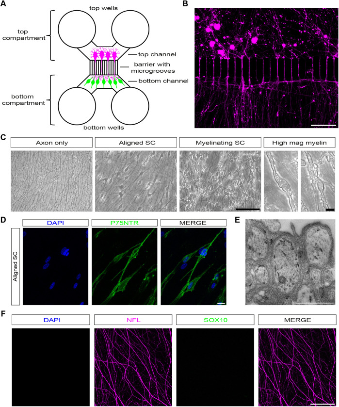
Myelinating Schwann cell (SC)-dorsal root ganglion (DRG) neuron cocultures are an important technique for understanding cell-cell signalling and interactions during peripheral nervous system (PNS) myelination, injury, and regeneration. Although methods using rat SCs and neurons or mouse DRG explants are commonplace, there are no established protocols for compartmentalised myelinating cocultures with dissociated mouse cells. There consequently is a need for a coculture protocol that allows separate genetic manipulation of mouse SCs or neurons, or use of cells from different transgenic animals to complement in vivo mouse experiments. However, inducing myelination of dissociated mouse SCs in culture is challenging. Here, we describe a new method to coculture dissociated mouse SCs and DRG neurons in microfluidic chambers and induce robust myelination. Cocultures can be axotomised to study injury and used for drug treatments, and cells can be lentivirally transduced for live imaging. We used this model to investigate axon degeneration after traumatic axotomy and find that SCs, irrespective of myelination status, are axo-protective. At later timepoints after injury, live imaging of cocultures shows that SCs break up, ingest and clear axonal debris.

Keywords: Axon degeneration; Coculture; Dorsal root ganglion neuron; Mouse; Myelination; Schwann cell; Wallerian degeneration.


Conflict of interest statement

Competing interests M.P.C. is a consultant for NuraBio. The remaining authors declare no competing interests.

Figures

Fig. 1.
Fig. 1.
Dissociated murine SC–DRG neuron cocultures in microfluidic chambers. (A) Standard neuron device with a 150 μm microgroove barrier. Dissociated DRG neurons (magenta), which cannot cross the barrier owing to their size, are seeded into the top channel and extend axons into the bottom channel, reaching the bottom wells. SCs (green) are then seeded in the bottom channel where they align with and myelinate axons. (B) Dissociated DRG neurons (magenta) growing across the microgroove barrier. Scale bar: 100 μm. (C) Phase images of the bottom channel showing axons only, axons with aligned SCs (aligned SC), and axons with myelinating SCs (myelinating SC). Scale bar: 100 μm. Higher magnification images show myelin segments in cultures with myelinating SCs. Scale bar: 10 μm. (D) Aligned SCs can be labelled with p75NTR. Scale bar: 20 μm. (E) Electron micrographs of aligned Schwann cells that show structures resembling Remak bundles. Scale bar: 1 nm. (F) Confocal images of axon-only cultures, showing axons (NFL, magenta), but no DAPI (blue) or SOX10 (green) signal in the axonal compartment of the microfluidic chamber. Scale bar: 100 μm. All images representative of at least three experimental repeats.
Fig. 2.
Fig. 2.
SCs in dissociated myelinating cocultures can be induced to robustly myelinate. (A) Confocal images of dissociated DRG neuron cocultures with aligned SC or myelinating SC. Axons are NFL-labelled (magenta), and SCs PRX-labelled (green). Aligned SCs have a more diffuse morphology (white arrowheads), while myelin segments are present in myelinating SCs (white arrows). Scale bar: 100 μm. (B) Higher magnification images showing NFL-labelled axons (magenta) covered by myelin segments (PRX, green, white arrows). Scale bar: 10 μm. (C) DRG neurons (NFL, magenta) growing across the barrier in chambers with SCs (PRX, green) that have been induced to myelinate. Scale bar: 100 μm. (D) Electron micrograph of electron-dense myelin in cocultures with myelinating SCs. Scale bar: 100 nm. (E) MPZ (green)-labelled myelinating SCs. Scale bar: 20 μm. (F) MBP (green)-labelled myelinating SCs. Scale bar: 10 μm. (G) In mature myelinating cultures, SCs CASPR (white) can be detected in the characteristic staining pattern marking paranodes. Scale bar 5 μm. Paranodal CASPR co-localised on axons (magenta). Scale bar: 5 μm. When colabelling with MPZ (green), CASPR is localised to paranodal loops adjacent to a node of Ranvier. Scale bar: 10 μm. All images representative of at least three experimental repeats.
Fig. 3.
Fig. 3.
SC–DRG neuron cocultures replicate characteristic axonal and SC injury responses after axotomy. (A) At 12 h post axotomy many of the axons (magenta) in SC–DRG neuron cocultures have degenerated. Scale bar: 100 μm. (B) SC JUN in uninjured cultures and 12 h post axotomy. With identical imaging conditions, a signal can only be detected 12 h post axotomy. Scale bar: 100 μm. (C) Relative intensity of JUN signal in uninjured myelinating cultures and 12 h post axotomy (n=3). Data shown in violin plots with median (line) and upper and lower quartiles (dotted lines) marked. (D) SC JUN in uninjured cultures with aligned Schwann cells and 12 h post axotomy. With identical imaging conditions, a signal can only be detected 12 h post axotomy. Scale bar 100 μm. (E) Relative intensity of JUN signal in uninjured cultures with aligned Schwann cells and 12 h post axotomy (n=3). Data shown in violin plots with median (line) and upper and lower quartiles (dotted lines) marked. Statistical analysis in C and E was performed with two-tailed unpaired Student's t-test. P values are shown in the figure. (F) Myelinating SCs demyelinate after extended periods of time (48 h) after axotomy. Myelin ovoids and myelin debris (both identified by white arrowheads) are present in fluoromyelin (green)-labelled cultures. Scale bar: 10 μm. (G) Electron micrographs of myelinating SCs at 48 h post axotomy showing characteristic demyelinated profiles surrounding degenerated axons. Scale bars: 1 μm. (H) Vacor induces neurodegeneration when applied to the top compartment of cocultures. NFL (magenta). Scale bar: 10 μm. (I) Neurons and SCs can be lentivirally infected prior to plating in microfluidic chambers. Neurons were infected with LV-CMV-mCherry (MOI 2, magenta) and SCs with LV-CMV-GFP (MOI 200, green). Scale bar: 20 μm. n number refers to independently prepared cultures from separate litters of mice. All images representative of at least three experimental repeats.
Fig. 4.
Fig. 4.
SCs are axo-protective independently of myelination status at early timepoints, and promote axon fragmentation at later timepoints after axotomy. (A) Confocal images of NFL-labelled cocultures prior to axotomy at the microgroove barrier (uninjured), and 3, 6, 9 and 12 h after axotomy. Axon-only cultures show earlier signs of degeneration than cultures with SCs. Scale bar: 100 μm. (B) Quantification of axon degeneration after axotomy. Only axon-only cultures show statistically substantial degeneration at 6 h post axotomy (29±1.44%, n=5). In axon-only cultures, this degeneration increases to 47.39±1.34% at 9 h post axotomy (n=3), and finally 76.68±6.60% at 12 h post axotomy (n=3). Cocultures with SCs show little degeneration at 3 h post axotomy (aligned SCs, 4.87±1.65%, n=3; myelinating SCs, 3.048±0.06%, n=3) and 6 h post axotomy (myelinating SCs, 8.14±0.65%, n=4; aligned SCs, 9.19±2.57%, n=3). At 9 and 12 h post axotomy, axons associated with both aligned and myelinating SCs start to degenerate (aligned SCs, 9 h, 30.05±3.05%, n=4 and 12 h, 44.61±4.72%, n=3; myelinating SCs, 9 h, 33.12±0.61%, n=3 and 12 h, 48.38±7.75%, n=3). There were no significant differences in axon degeneration rates between aligned and myelinating SCs cocultures (3 h, P=0.65; 6 h, P=0.98; 9 h, P=0.44; 12 h, P=0.70). Axon-only cultures show significant differences compared to cultures with aligned or myelinating SCs at 6 h (aligned SCs, P<0.0001; myelinating SCs, P<0.0001), 9 h (aligned SCs, P=0.0004; myelinating SC, P=0.0059), and 12 h (aligned SCs, P<0.0001, myelinating SC: P<0.0001) post axotomy. Results shown as individual data points. Statistical significance for comparisons between axon-only cultures and aligned SC cocultures are displayed on the graph. ***P<0.001; ****P<0.0001; ns, not significant. All values are mean±s.e.m.; n number refers to independently prepared cultures from separate litters of mice. Statistical analysis was performed with two-way ANOVA with post-hoc Tukey test to correct for multiple comparisons. (C) Confocal images of mCherry-labelled axons (magenta) and GFP-labelled SCs (green). At an early timepoint (19 h 10 min) an intact axon is visible (white arrow); 3 h later (22 h 10 min), the axon is starting to be constricted (white arrowheads). This continues until 23 h 50 min after axotomy, when constrictions are clearly visible, and the axon is swollen between two constrictions. At 24 h after axotomy, the axon then breaks apart. Scale bar: 10 μm. (D) Confocal images of mCherry-labelled axons (magenta) and GFP-labelled SCs (green) with intact axons (white arrow, 28 h post axotomy) just before axon degeneration and with mCherry fragments (white arrowheads) within SCs 30 h post axotomy, once axon degeneration has occurred. Scale bar: 10 μm. All images representative of at least three experimental repeats for studies of degeneration rates (A,B) and two experimental repeats for live imaging of axon fragmentation (C,D).

Similar articles

Cited by

References

    1. Arthur-Farraj, P. and Coleman, M. P. (2021). Lessons from injury: how nerve injury studies reveal basic biological mechanisms and therapeutic opportunities for peripheral nerve diseases. Neurotherapeutics 18, 2200-2221. 10.1007/s13311-021-01125-3 - DOI - PMC - PubMed
    1. Arthur-Farraj, P., Wanek, K., Hantke, J., Davis, C. M., Jayakar, A., Parkinson, D. B., Mirsky, R. and Jessen, K. R. (2011). Mouse Schwann cells need both NRG1 and cyclic AMP to myelinate. Glia 59, 720-733. 10.1002/glia.21144 - DOI - PMC - PubMed
    1. Arthur-Farraj, P. J., Latouche, M., Wilton, D. K., Quintes, S., Chabrol, E., Banerjee, A., Woodhoo, A., Jenkins, B., Rahman, M., Turmaine, M.et al. (2012). c-Jun reprograms Schwann cells of injured nerves to generate a repair cell essential for regeneration. Neuron 75, 633-647. 10.1016/j.neuron.2012.06.021 - DOI - PMC - PubMed
    1. Arthur-Farraj, P. J., Morgan, C. C., Adamowicz, M., Gomez-Sanchez, J. A., Fazal, S. V., Beucher, A., Razzaghi, B., Mirsky, R., Jessen, K. R. and Aitman, T. J. (2017). Changes in the coding and non-coding transcriptome and DNA methylome that define the Schwann cell repair phenotype after nerve injury. Cell Rep. 20, 2719-2734. 10.1016/j.celrep.2017.08.064 - DOI - PMC - PubMed
    1. Babetto, E., Wong, K. M. and Beirowski, B. (2020). A glycolytic shift in Schwann cells supports injured axons. Nat. Neurosci. 23, 1215-1228. 10.1038/s41593-020-0689-4 - DOI - PMC - PubMed

Publication types