. 2023 Oct 1;325(4):L487-L499.
doi: 10.1152/ajplung.00227.2022. Epub 2023 Aug 29.

Investigating the role of platelets and platelet-derived transforming growth factor-β in idiopathic pulmonary fibrosis

Affiliations

Investigating the role of platelets and platelet-derived transforming growth factor-β in idiopathic pulmonary fibrosis

Deborah L W Chong et al. Am J Physiol Lung Cell Mol Physiol. .

Abstract

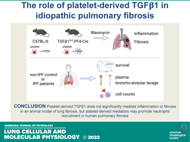
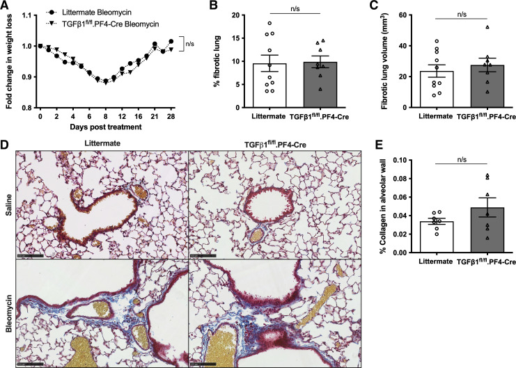
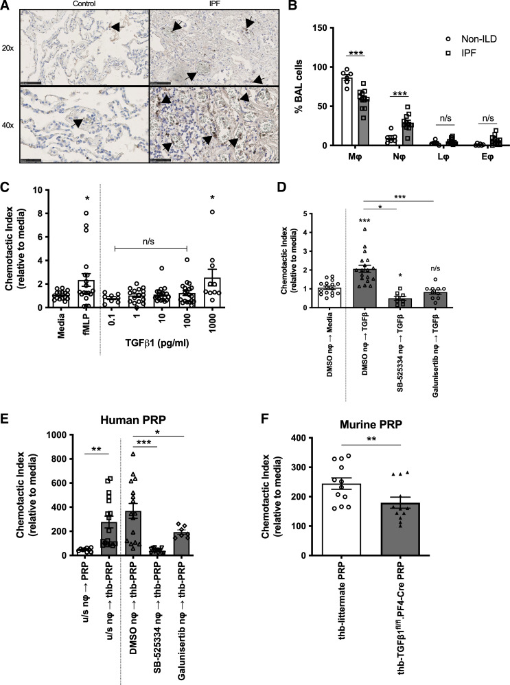
Transforming growth factor-β1 (TGFβ1) is the key profibrotic cytokine in idiopathic pulmonary fibrosis (IPF), but the primary source of this cytokine in this disease is unknown. Platelets have abundant stores of TGFβ1, although the role of these cells in IPF is ill-defined. In this study, we investigated whether platelets, and specifically platelet-derived TGFβ1, mediate IPF disease progression. Patients with IPF and non-IPF patients were recruited to determine platelet reactivity, and separate cohorts of patients with IPF were followed for mortality. To study whether platelet-derived TGFβ1 modulates pulmonary fibrosis (PF), mice with a targeted deletion of TGFβ1 in megakaryocytes and platelets (TGFβ1fl/fl.PF4-Cre) were used in the well-characterized bleomycin-induced pulmonary fibrosis (PF) animal model. In a discovery cohort, we found significantly higher mortality in patients with IPF who had elevated platelet counts within the normal range. However, our validation cohort did not confirm this observation, despite significantly increased platelets, neutrophils, active TGFβ1, and CCL5, a chemokine produced by inflammatory cells, in the blood, lung, and bronchoalveolar lavage (BAL) of patients with IPF. In vivo, we showed that despite platelets being readily detected within the lungs of bleomycin-treated mice, neither the degree of pulmonary inflammation nor fibrosis was significantly different between TGFβ1fl/fl.PF4-Cre and control mice. Our results demonstrate for the first time that platelet-derived TGFβ1 does not significantly mediate inflammation or fibrosis in a PF animal model. Furthermore, our human studies revealed blood platelet counts do not consistently predict mortality in IPF but other platelet-derived mediators, such as C-C chemokine ligand 5 (CCL5), may promote neutrophil recruitment and human IPF.NEW & NOTEWORTHY Platelets are a rich source of profibrotic TGFβ; however, the role of platelets in idiopathic pulmonary fibrosis (IPF) is unclear. We identified that patients with IPF have significantly more platelets, neutrophils, and active TGFβ in their airways than control patients. Using an animal model of IPF, we demonstrated that platelet-derived TGFβ does not significantly drive lung fibrosis or inflammation. Our findings offer a better understanding of platelets in both human and animal studies of IPF.

Keywords: inflammation; interstitial lung diseases; platelets; pulmonary fibrosis; transforming growth factor-β1.


Conflict of interest statement

No conflicts of interest, financial or otherwise, are declared by the authors.

Figures

None
Graphical abstract
Figure 1.
Figure 1.
Human blood platelet counts do not consistently predict mortality in IPF. A and B: Kaplan–Meier survival curves of patients with IPF from two different cohorts: UCLH discovery cohort (n = 214 patients; A) or Exeter validation cohort of patients with IPF (n = 285 patients; B). Patients were divided into three strata based on blood platelet count (see Supplemental Methods for blood cutoff values for tertiles). The three strata were defined bias-free by dividing into three equal groups: lowest tertile (blue), middle tertile (red), and highest tertile (green). C: blood platelet counts of patients with IPF (n = 162) or healthy controls (n = 3,77,093) from UK Biobank data. Means ± SD are shown. Any statistical differences between survival curves or blood platelet counts were determined using the Gehan–Breslow–Wilcoxon test or unpaired t test (**P < 0.01). IPF, idiopathic pulmonary fibrosis.
Figure 2.
Figure 2.
Levels of active TGFβ1 correlate with an activated blood platelet signature in patients with IPF. Concentrations of total (A) or active TGFβ1 (B), CXCL4 (C), P-Selectin (D), and MMP7 (E) were measured in the plasma of patients with non-ILD and IPF (n = 10 and 37, respectively) by ELISA or MLEC bioassay. Concentrations of total (F) or active TGFβ1 (G) or MMP7 (H) in the plasma of patients with IPF were correlated with paired blood platelet counts (n = 33). Concentrations of MMP7 (I), total (J), or active TGFβ1 (K) in the plasma of patients with IPF were correlated with paired P-Selectin levels (n = 37). Concentrations of MMP7 (L), total (M), or active (N) TGFβ1 in the plasma of patients with IPF were correlated with paired CXCL4 levels (n = 37). Any statistical differences were determined using unpaired t test or linear regression and any significant differences are indicated (n/s = not significant, *P < 0.05, **P < 0.01. IPF, idiopathic pulmonary fibrosis; MLEC, Mink lung epithelial cell bioassay; non-ILD, non-interstitial lung diseases.
Figure 3.
Figure 3.
Platelet-derived TGFβ1 does not play a significant role in bleomycin-induced fibrosis. A: quantification of secreted active TGFβ1 by MLEC bioassay in unstimulated (white bars) or thrombin-treated (gray bars) PRP from littermate (n = 4) or TGFβ1fl/fl.PF4-Cre (n = 5) mice. B: TGFβ1fl/fl.PF4-Cre or littermate mice were given 50 IU bleomycin (n = 5/group) or saline (n = 3/group) via an oropharyngeal route. Fold-change in body weight loss was monitored for 21 days post instillation. C: representative micro-CT scans of lungs after 21 days of treatment (white asterisks denote fibrotic lesions). Percentage (D) or volume (E) of fibrotic lung tissue in micro-CT scanned lungs was determined using InForm analysis software (n = 3 saline/mouse group and n = 5 bleomycin/mouse group). F: total lung collagen of murine lungs was determined by reverse-phase HPLC (n = 3 saline/mouse group and n = 5 bleomycin/mouse group). Any statistical differences were determined using two-way ANOVA with Holm–Sidak post hoc testing or Mann–Whitney U tests. In B, asterisks represent significant differences between treatments in littermate control mice and apostrophes represent significant differences between treatments in TGFβ1fl/fl.PF4-Cre mice. In D and E, asterisks above the bars represent significance between saline and bleomycin treatment. Any other significant differences are indicated (n/s = not significant, *P < 0.05, **P < 0.01, ***P < 0.001). MLEC, Mink lung epithelial cell bioassay; PRP, platelet-rich plasma.
Figure 4.
Figure 4.
Platelet-derived TGFβ1 does not play a significant role in disease resolution after bleomycin-induced injury. A: TGFβ1fl/fl.PF4-Cre (n = 8) or littermate mice (n = 10) were given 25 IU bleomycin via an oropharyngeal route. Fold-change in body weight loss was monitored for 28 days post instillation. Percentage (B) and volume (C) of fibrotic lung tissue in micro-CT scanned lungs were determined using InForm analysis software (n = 10 littermate controls or n = 8 TGFβ1fl/fl.PF4-Cre mice). D: representative images of modified Martius Scarlet Blue (MSB) trichrome stained saline- (top) or bleomycin-treated (bottom) littermate control (left) or TGFβ1fl/fl.PF4-Cre (right) lung sections from two independent experiments (blue = collagen, yellow = RBC, red = cytoplasm, dark red = nuclei). Images are shown as ×20 magnification as denoted by 100 µm scale bar. E: quantification of percentage of alveolar collagen deposition in bleomycin-treated littermate control or TGFβ1fl/fl.PF4-Cre lungs based on MSB stained lungs. No statistical differences were found using two-way ANOVA with Holm–Sidak post hoc testing or Mann–Whitney U tests (n/s = not significant).
Figure 5.
Figure 5.
Platelet-derived TGFβ1 partially contributes to neutrophil chemotaxis. A: CD61+ platelets and platelet aggregates (indicated by the arrows) in control and IPF lung were detected by IHC. Size denoted by scale bar (×20 scale bar = 100 µm, ×4 scale bar = 50 µm). B: cell counts of macrophages (mϕ), neutrophils (nϕ), lymphocytes (lϕ), and eosinophils (eϕ) were determined in patients with non-ILD (n = 6) and IPF (n = 11) by cytospins of recovered cells in BALF. C: chemotaxis of human neutrophils (n = 4 healthy donors) toward media, 100 nM fMLP or increasing TGFβ1 concentrations (0.1–1,000 pg/mL). D: chemotaxis of DMSO, SB-525334 or Galunisertib ALK5 inhibitors pretreated human peripheral neutrophils (n = 3 or 4 healthy donors) toward media (white bar) or 1 ng/mL TGFβ1 (dark gray bars). E: chemotaxis of untreated, DMSO, SB-525334 or Galunisertib ALK5 inhibitors pretreated human peripheral neutrophils (n = 3 or 4 healthy donors) toward unstimulated (white bar) or thrombin (thb)-activated human PRP (gray bars). F: chemotaxis of murine bone marrow-derived neutrophils (n = 4 littermate mice) toward thrombin (thb)-activated littermate (white bar) or TGFβ1fl/fl.PF4-Cre (black bar) murine PRP. Statistical differences were determined using Mann–Whitney U test or one-way ANOVA with Holm–Sidak post hoc testing (n/s = not significant, *P < 0.05, **P < 0.01, ***P < 0.001). IHC, immunohistochemistry; IPF, idiopathic pulmonary fibrosis; non-ILD, non-interstitial lung diseases; PRP, platelet-rich plasma.
Figure 6.
Figure 6.
Platelet-derived TGFβ1 does not contribute to neutrophil recruitment during bleomycin-induced inflammation. A: TGFβ1fl/fl.PF4-Cre or littermates were given 50 IU bleomycin or saline (n = 9 littermate saline, n = 4 TGFβ1fl/fl.PF4-Cre saline, n = 15 littermate bleomycin, n = 8 TGFβ1fl/fl.PF4-Cre bleomycin) via an oropharyngeal route. Fold-change in body weight loss was monitored for 6 days post instillation. B: representative BALF cytospins after 6 days of treatment. Size denoted by 100-µm scale bar. Neutrophil (C), macrophages (D), or lymphocyte (E) populations from recovered BALF were quantified from cytospins (n = 9 littermate saline, n = 4 TGFβ1fl/fl.PF4-Cre saline, n = 15 littermate bleomycin, n = 8 TGFβ1fl/fl.PF4-Cre bleomycin). F: representative flow cytometric gating strategy to distinguish recovered cell populations from lung homogenate. Neutrophil (G), alveolar (H), or inflammatory macrophage cell (I) populations in lung homogenate were quantified by flow cytometry (n = 9 littermate saline, n = 4 TGFβ1fl/fl.PF4-Cre saline, n = 15 littermate bleomycin, n = 8 TGFβ1fl/fl.PF4-Cre bleomycin). Statistical differences were determined using one-way or two-way ANOVA with Holm–Sidak post hoc testing. In A, asterisks represent significance between treatments for littermate mice and apostrophes represent significance between treatments for TGFβ1fl/fl.PF4-Cre mice. In C and E, asterisks above the bars represent significance between saline or bleomycin group. Any other significant differences are indicated (n/s = not significant, *P < 0.05, **P < 0.01, ***P < 0.001). BALF, bronchoalveolar lavage fluid; IPF, idiopathic pulmonary fibrosis.
Figure 7.
Figure 7.
Platelet-derived mediators may contribute to neutrophil recruitment in IPF lung and disease severity. A: representative flow cytometric plot showing CD61 expression in platelet gate in IPF BALF. B: percentage of CD61+ platelets of total cells in BALF was quantified by flow cytometric analysis (n = 3 non-ILD and n = 5 IPF). MMP7 (C), total (D) or active TGFβ1 (E), CXCL4 (F) or CCL5 (G) concentrations were measured in BALF of patients with non-ILD and IPF (n = 7 and 16, respectively) by ELISA or MLEC bioassay. Concentrations of MMP7 (H), total (I) or active TGFβ1 (J), or CCL5 (K) in BALF of patients with IPF were correlated with paired CXCL4 (n = 16). L: concentrations of CCL5 in BALF of patients with IPF were correlated with paired MMP7 (n = 16). Statistical differences were determined using Mann–Whitney U tests or linear regression and any significant differences are indicated (n/s = not significant, *P < 0.05, **P < 0.01, ***P < 0.001). BALF, bronchoalveolar lavage fluid; IPF, idiopathic pulmonary fibrosis; MLEC, Mink lung epithelial cell bioassay; non-ILD, non-interstitial lung diseases.

References

    1. Mikolasch TA, Garthwaite HS, Porter JC. Update in diagnosis and management of interstitial lung disease. Clin Med (Lond) 16: s71–s78, 2016. doi:10.7861/clinmedicine.16-6-s71. - DOI - PMC - PubMed
    1. Case AH. Clinical overview of progressive fibrotic interstitial lung disease. Front Med (Lausanne) 9: 858339, 2022. doi:10.3389/fmed.2022.858339. - DOI - PMC - PubMed
    1. Mei Q, Liu Z, Zuo H, Yang Z, Qu J. Idiopathic pulmonary fibrosis: an update on pathogenesis. Front Pharmac 12: 797292, 2022. doi:10.3389/fphar.2021.797292. - DOI - PMC - PubMed
    1. Moss BJ, Ryter SW, Rosas IO. Pathogenic mechanisms underlying idiopathic pulmonary fibrosis. Annu Rev Pathol 17: 515–546, 2022. doi:10.1146/annurev-pathol-042320-030240. - DOI - PubMed
    1. Karimi-Shah BA, Chowdhury BA. Forced vital capacity in idiopathic pulmonary fibrosis — FDA review of pirfenidone and nintedanib. N Engl J Med 372: 1189–1191, 2015. doi:10.1056/NEJMp1500526. - DOI - PubMed

Publication types

Substances

LinkOut - more resources