. 2023 Oct 9;8(19):e170928.
doi: 10.1172/jci.insight.170928.

Acid-base homeostasis orchestrated by NHE1 defines the pancreatic stellate cell phenotype in pancreatic cancer

Affiliations

Acid-base homeostasis orchestrated by NHE1 defines the pancreatic stellate cell phenotype in pancreatic cancer

Zoltán Pethő et al. JCI Insight. .

Abstract

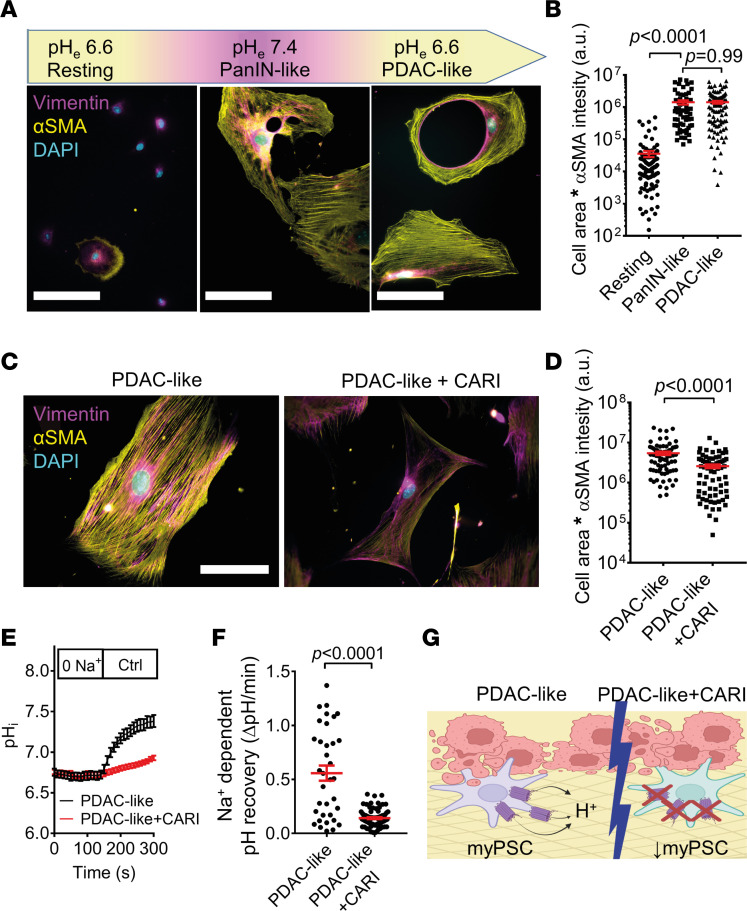
Pancreatic ductal adenocarcinoma (PDAC) progresses in an organ with a unique pH landscape, where the stroma acidifies after each meal. We hypothesized that disrupting this pH landscape during PDAC progression triggers pancreatic stellate cells (PSCs) and cancer-associated fibroblasts (CAFs) to induce PDAC fibrosis. We revealed that alkaline environmental pH was sufficient to induce PSC differentiation to a myofibroblastic phenotype. We then mechanistically dissected this finding, focusing on the involvement of the Na+/H+ exchanger NHE1. Perturbing cellular pH homeostasis by inhibiting NHE1 with cariporide partially altered the myofibroblastic PSC phenotype. To show the relevance of this finding in vivo, we targeted NHE1 in murine PDAC (KPfC). Indeed, tumor fibrosis decreased when mice received the NHE1-inhibitor cariporide in addition to gemcitabine treatment. Moreover, the tumor immune infiltrate shifted from granulocyte rich to more lymphocytic. Taken together, our study provides mechanistic evidence on how the pancreatic pH landscape shapes pancreatic cancer through tuning PSC differentiation.

Keywords: Epithelial transport of ions and water; Fibrosis; Homeostasis; Metabolism; Oncology.


Conflict of interest statement

Conflict of interest: The authors have declared that no conflict of interest exists.

Figures

Figure 1
Figure 1. Environmental alkalization induces myofibroblastic PSC differentiation and proliferation.
(A) Concept of the working hypothesis. In the healthy pancreas, the marked HCO3 secretion upon each meal results in a distinct stromal acidification, keeping the PSCs in a quiescent nonfibrotic phenotype. Upon malignant transformation in early PDAC (PanIN), ductal secretion decreases, resulting in a relief of the intermittent acidity ( = relative stromal alkalization), leading to a myofibroblastic PSC differentiation. Acidic → alkaline pHe is depicted by yellow → purple colors. (B) Hallmark gene set enrichment analysis (GSEA) of RNA-Seq data from PSCs cultured at pHe7.4 versus pHe6.6 shows the top 5 differentially regulated pathways (n/N = 3/3). (C) A heatmap of RNA-Seq expression mean Z scores computed for published signature genes of immunomodulatory CAFs (left) and myofibroblastic CAFs (right). The gene (rows) Z scores for pHe6.6 and pHe7.4 are color coded. Dark green indicates higher expression Z scores (n/N = 3/3). (D) Immunocytochemistry images of PSCs cultured at pHe6.6 or pHe7.4. The activation marker αSMA (yellow) and the general PSC marker vimentin (magenta) are labeled. Nuclei are stained with DAPI (cyan). Scale bar: 50 μm. (E) Cell areas multiplied by αSMA intensity is taken as a readout for the myofibroblastic PSC phenotype. It is plotted as a function of pHe. Mean values are shown as n ≥ 142 from N = 3 mice. Half-maximum (EC50) pHe-dependent PSC activation occurs at pHe7.0. Note the logarithmic scale of the ordinate. (F) Representative Western blot (top) of p53 and GAPDH of PSCs cultured at pHe6.6 or pHe7.4, with subsequent quantification (bottom) (n/N = 3/3). (G) Representative cell cycle histogram of PSCs cultured at pHe6.6 (red) and pHe7.4 (blue), assessed by flow cytometry with propidium iodide (PI) staining. Cell populations at different stages of the cell cycle are indicated by arrows. (H) The bar chart depicts the percentage of PSCs at the G0/G1 phase of the cell cycle when cultured at pHe 6.6 (red) and pHe7.4 (blue). Data points are npHe6.6 = 3 and npHe7.4 = 5 measurements from n = 3 individual mice. Statistical tests in F and H were performed with 2-tailed unpaired Student’s t tests. A was created with BioRender.com.
Figure 2
Figure 2. NHE1-mediated pH recovery is inhibited by cariporide in PSCs.
(A) Volcano plot analysis of ion transporter genes (GO:0015075) derived from the RNA-Seq data of PSCs cultured at pHe7.4 and pHe6.6 (n/N = 3/3). Genes indicated by red (n = 43) and blue (n = 44) dots highlighted in rectangles are upregulated at pHe6.6 and pHe7.4, respectively. (B) Subsequent qPCR validation of ion transporter gene expression levels by the 2–ΔCT method compared with the housekeeping genes Ywhaz and 18s rRNA. Bar charts show mean expression of genes from freshly isolated quiescent PSCs (0 hours, white) and PSCs cultured at pHe7.4 (blue) or pHe6.6 (red) (n/N = 6/3). (C) Representative immunofluorescence images showing the cellular localization of NHE1 in freshly isolated quiescent PSCs (0 hours), PSCs cultured at pHe7.4 or pHe6.6 for 120 hours, or vehicle-treated KPfC-derived PSCs (PDAC-PSC) (NHE1: magenta; DAPI: blue). Scale bar: 10 μm. (D) NHE1 Western blots are shown, with the top bands at 100 kDa corresponding to the glycosylated NHE1, whereas bands with lower molecular weight (80 kDa) correspond to the unglycosylated NHE1. Lysates are from N = 3 mice each. (E) pHi recordings of WT PSCs cultured at pHe6.6 (left) and pHe7.4 (middle) or KPfC-derived CAFs (cultured at pHe7.4; right). pHi was acidified temporarily by applying the NH4+ prepulse (*) technique. NHE1-independent pHi recovery starts when pHi has reached its minimum in the presence of the Na+-free solution (“0 Na+”). NHE1-dependent pHi recovery can be observed in the last step (Ctrl) of the experiment when cariporide was added to the Na+-containing superfusion as indicated. Lines show mean pHi of npH6.6 = 35, npH6.6+CARI = 39, npH7.4 = 45, npH7.4+CARI = 68, nPDAC-CAF = 11, and nPDAC-CAF+CARI = 22 cells from N = 3 mice each. (F) Quantification of resting pHi of PSCs cultured at pHe6.6 (red) or pHe7.4 (blue) and CAFs (purple), respectively, derived from E (npH6.6 = 74, npH7.4 = 113, and nPDAC-CAF+CARI = 41 cells from N = 3 mice each). (G) Scatter plot depicts the rate of Na+-independent pHi recovery of PSCs cultured at pHe6.6 (red), pHe7.4 (blue), or CAFs (purple), derived from E (n/N see F). (H) Comparison of the rate of Na+-dependent pHi recovery of WT PSCs cultured at pHe6.6 (red) or pHe7.4 (blue), or CAFs (purple) as explained in E (n/N see E). Statistical tests in FH were performed with 1-way ANOVA with Tukey’s post hoc test.
Figure 3
Figure 3. PSC mechanotransduction mediated by YAP1 is inhibited at acidic pHe.
(A) Immunocytochemistry images of PSCs cultured on substrates of different stiffnesses at pHe6.6 (top) and pHe7.4 (bottom). The activation marker αSMA (yellow) and the general stellate cell marker vimentin (magenta), as well as nuclei (cyan), are labeled. Scale bar: 50 μm. (B) Cell area multiplied with mean αSMA fluorescence intensity was taken as a readout of myofibroblastic PSC phenotype on hydrogels with 11 kPa stiffness or on glass (1 GPa). n11kPa = 65, n1GPa = 170 cells from N = 3 mice. (C) Representative immunofluorescence images of YAP1 in PSCs (green) under the indicated cell culture conditions. YAP1, when translocated from the cytosol (#) into the nucleus (*), initiates transcription. Scale bar: 50 μm. (D) The ratio of nuclear/cytosolic YAP1 fluorescence intensity was determined as a readout of YAP1-mediated signal transduction. n11kPa-pH6.6 = 68, n11kPa-pH7.4 = 51, n1GPa-pH6.6 = 43, n1GPa+pH7.4 = 63 cells from N = 3 mice each. Statistical tests in B and D were performed with 1-way ANOVA with Tukey’s post hoc test.
Figure 4
Figure 4. The myofibroblastic phenotype of activated PSCs is partially reversed by cariporide but not by acidic pHe alone.
(A) After culturing PSCs at pHe6.6 for 72 hours, medium was changed to pHe6.6 (Resting) or to pHe7.4 (PanIN-like) for another 72 hours. Lastly, the medium of pHe7.4-incubated cells was reacidified to pHe6.6 for another 72 hours (PDAC-like). Representative images of PSCs stained for αSMA (yellow), vimentin (magenta), and DAPI (cyan) are shown below each condition. Scale bar: 50 μm. (B) Scatter plot shows cell areas multiplied by αSMA fluorescence staining intensity (logarithmic scale) under conditions described in A. nResting = 100, nPanIN-like = 64, and nPDAC-like = 94; N = 3 mice. (C) Immunocytochemistry of PDAC-like PSCs in the absence (left) or presence (right) of cariporide (CARI). Scale bar: 50 μm. (D) Scatter plot shows cell areas multiplied by αSMA fluorescence intensity (logarithmic scale) under conditions described in C. nPDAC-like = 74, nPDAC-like+CARI = 74; N = 3 mice. (E) Intracellular pH measurements of PSCs where culture medium is reacidified after activation (pHe7.4 → pHe6.6, PDAC-like). Intracellular pH was acidified temporarily by applying the NH4+ prepulse technique, as shown in Figure 2. NHE1-dependent pH recovery can be observed when cells are superfused with Na+-containing solution (Ctrl) without (black) or with cariporide (red). Lines indicate the mean pHi of nPDAC-like = 35, nPDAC-like+CARI = 79 cells from N = 3 mice. (F) Comparison of the rate of Na+-dependent recovery of PSCs in the absence or presence of cariporide derived from E. (G) Illustration of extended working hypothesis. In manifest PDAC, acidic pHe fails to acidify pHi because of NHE1-mediated H+ extrusion (left). Therefore, PSCs and CAFs remain myofibroblastic, ultimately promoting tumor desmoplasia. However, upon NHE1 inhibition with cariporide, PSCs and CAFs fail to counterbalance the acid stress, which disrupts the myofibroblastic phenotype (right). Statistical comparison in B was performed with 1-way ANOVA with Tukey’s post hoc test, whereas in D and F, statistical comparisons were performed with 2-tailed unpaired Student’s t tests. G was created with BioRender.com.
Figure 5
Figure 5. NHE1 inhibitor treatment leads to reduced desmoplastic reaction in murine PDAC.
(A) Schematic representation of the 4-week–long treatment protocol of KPfC mice. Treatment started at the age of week 20. Cariporide was applied daily (1/D), and gemcitabine (100 mg/kg i.p.) was coinjected with cariporide (3 mg/kg i.p.) on the days indicated by the arrows. (B) Total pancreas volume of KPfC mice after gemcitabine (GEM) or cariporide (CARI) monotherapy or gemcitabine + cariporide (GEM+CARI) combined chemotherapy. Inlet demonstrates that pancreas volume was measured via volume displacement. Data points depict individual pancreata; NVehicle = 11, NGEM = 9, NCARI = 11, NGEM+CARI = 11. (C) Relative tumor area in histological KPfC tissue sections was obtained by dividing total tumor area by total tissue area after H&E staining. Data points depict individual pancreata; NVehicle = 11, NGEM = 9, NCARI = 11, NGEM+CARI = 11. (D) Representative images of PDAC nodes (marked with *) after H&E and Sirius red stainings. The degree of fibrosis correlates with the area of Sirius red+ (marked in red, #) tissue neighboring the cancerous tissue. Scale bar: 100 μm. (E) Relative tumor fibrosis of each Sirius red–stained KPfC tissue section was determined by dividing the summed area of fibrosis within every tumor node (sum of thresholded black areas in every node in the inlet) by the summed area of the tumor nodes. Data points depict individual pancreata; NVehicle = 11, NGEM = 9, NCARI = 11, NGEM+CARI = 12. (F) To obtain the fibrosis per tumor node, the area of Sirius red+ fibrosis (black thresholded area in the inlet) was divided by the total area of the respective tumor node. Data points depict individual tumor nodes; nVehicle = 400, nGEM = 239, nCARI = 279, nGEM+CARI = 476. Data and statistical comparison in B, C, and F are shown as median ± 95% CI with Kruskal-Wallis statistical test with Dunn’s post hoc test, and in E as mean ± SEM with 1-way ANOVA with Tukey’s post hoc test. Inlets for A and B were created with BioRender.com.
Figure 6
Figure 6. NHE1 orchestrates PDAC-derived CAF activation.
(A) Representative H&E and IHC images of healthy and tumorous ducts. The colors of the IHC image indicate cell nuclei stained with NHE1 (magenta), αSMA+ PSCs and CAFs (yellow), DAPI (cyan), and CK19+ ductal or tumor cells (green). Scale bar: 40 μm. (B) αSMA+ cells (*) from A are depicted with higher magnification, which are also NHE1+. Scale bar: 20 μm (C) Immunocytochemistry of CAFs derived from KPfC mice after a 1-month treatment with vehicle (top) or gemcitabine + cariporide (bottom). Myofibroblast marker αSMA (yellow), the general mesenchymal marker vimentin (magenta), and nuclei (cyan) are labeled. Scale bar: 20 μm. (D) KPfC-derived CAF activation after therapy was assessed by multiplying cell area with the fluorescence intensity of αSMA. nVehicle = 61, nGEM = 59, nCARI = 63, nGEM+CARI = 70 from N ≥ 3 mice. Note the logarithmic scale of the ordinate. (E) Trajectories of migrating KPfC-derived CAFs are shown by individual black lines. The treatment of the respective mice is indicated. Trajectories of the treatment groups are always normalized to common starting points. The radii of the orange circles highlight the mean translocation of cells in each population. Scale bar: 20 μm. (F) Mean cell migration velocities of individual CAFs were calculated from the trajectories in C. nVehicle = 30, nGEM = 30, nCARI = 19, nGEM+CARI = 40 cells from N ≥ 3 mice. Statistical tests in D and F were performed with 1-way ANOVA with Tukey’s post hoc test.
Figure 7
Figure 7. Lymphocyte/neutrophil ratio increases upon NHE1 inhibition in tumor sections of KPfC mice.
(A) H&E (left) and PAS-stained KPfC mouse tissue sections after vehicle and gemcitabine + cariporide (GEM+CARI) therapy. Cells of innate immunity, such as neutrophils (arrows), utilize glycogen and are thus PAS+ (purple), in contrast to, for example, lymphocytes. Scale bar: 50 μm. (B) Representative IHC images stained for Ly6G+ neutrophils (magenta, arrows on left image), CD3+ lymphocytes (yellow, arrows on the right image), and nuclei with DAPI (cyan). Scale bar: 50 μm. (C) CD3/Ly6G ratio was assessed by dividing the number of CD3+ cells by the number of Ly6G+ cells in every tumor node. Data points depict the mean CD3/Ly6G ratio derived from each tumor node in individual mice; NVehicle = 10, NGEM = 9, NCARI = 10, NGEM+CARI = 11 mice. (D) To obtain the CD3/Ly6G ratio per tumor node, the number of CD3+ cells was divided by the respective number of Ly6G+ cells in each tumor node. Data points depict individual tumor nodes; nVehicle = 386, nGEM = 276, nCARI = 301, nGEM+CARI = 398. Data and statistical comparison in D and E are represented as median ± 95% CI using Kruskal-Wallis statistical test with Dunn’s post hoc test.

Similar articles

Cited by

References

    1. American Cancer Society. Cancer Facts & Figures 2023. https://www.cancer.org/content/dam/cancer-org/research/cancer-facts-and-... Accessed August 30, 2023.
    1. Amrutkar M, Gladhaug IP. Stellate cells aid growth-permissive metabolic reprogramming and promote gemcitabine chemoresistance in pancreatic cancer. Cancers (Basel) 2021;13(4):601. doi: 10.3390/cancers13040601. - DOI - PMC - PubMed
    1. Cruz-Monserrate Z, et al. Targeting pancreatic ductal adenocarcinoma acidic microenvironment. Sci Rep. 2014;4:4410. doi: 10.1038/srep04410. - DOI - PMC - PubMed
    1. Hofschröer V, et al. Ion channels orchestrate pancreatic ductal adenocarcinoma progression and therapy. Front Immunol. 2021;11:586599. doi: 10.3389/fphar.2020.586599. - DOI - PMC - PubMed
    1. Wörmann SM, et al. The immune network in pancreatic cancer development and progression. Oncogene. 2014;33(23):2956–2967. doi: 10.1038/onc.2013.257. - DOI - PubMed

Publication types