

MACULAR NEUROVASCULAR ABNORMALITIES IN A CHILD WITH INCONTINENTIA PIGMENTI ON HANDHELD OPTICAL COHERENCE TOMOGRAPHY ANGIOGRAPHY
- PMID: 37643049
- PMCID: PMC12255511
- DOI: 10.1097/ICB.0000000000001264
MACULAR NEUROVASCULAR ABNORMALITIES IN A CHILD WITH INCONTINENTIA PIGMENTI ON HANDHELD OPTICAL COHERENCE TOMOGRAPHY ANGIOGRAPHY
Abstract
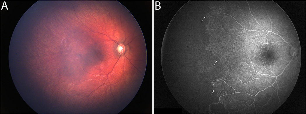
Purpose: To report macular neurovascular abnormalities in a child with incontinentia pigmenti using handheld optical coherence tomography (OCT) and OCT angiography (OCT-A).
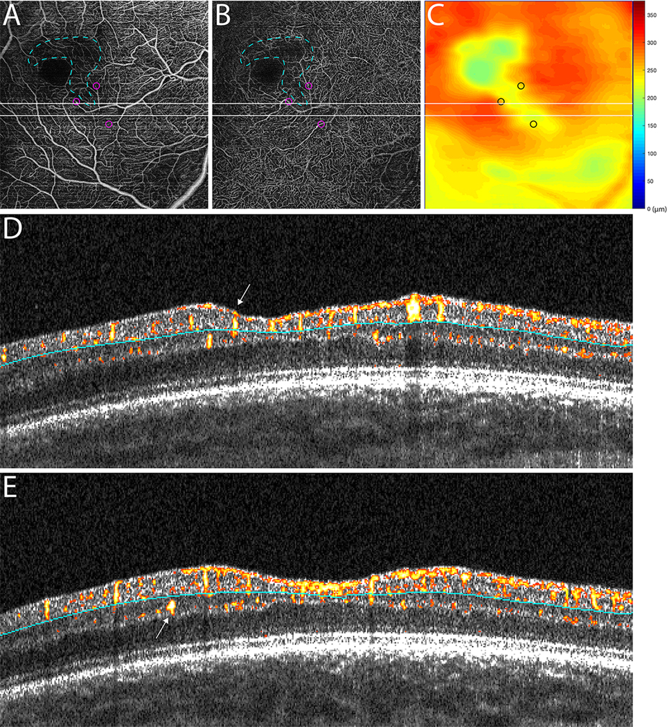
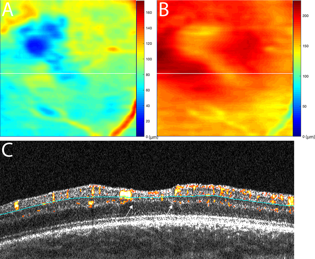
Methods: An eye of a child with incontinentia pigmenti enrolled in BabySTEPS was imaged using an investigational noncontact, handheld swept-source OCT device during examination under anesthesia. Custom MATLAB scripts were used to generate depth-resolved vascular slabs, B-scans with flow overlay, and retinal thickness maps.
Results: Depth-resolved OCT and OCT-A imaging demonstrated focal areas of decreased capillary flow that corresponded to areas of both inner retinal and outer retinal thinning on retinal thickness maps. Atypical diving of superficial retinal vessels occurred as they traversed from thin retina to normal-thickness retina.
Conclusion: Depth-resolved OCT and OCT-A identified retinal vascular abnormalities that were not evident on fundus photography or fluorescein angiography. This case depicted concurrent, localized abnormalities in retinal thickness and microvasculature in an eye with incontinentia pigmenti.
Conflict of interest statement
J. A. Izatt and C. A. Toth have a segmentation related patent (US10366492B2). C. Viehland, J. A. Izatt, and C. A. Toth and Duke University have a patent application pending on the novel handheld probe described in this manuscript. C. A. Toth receives royalties through her university, from Alcon. C. A. Toth consults for EMMES Inc. C. Viehland and C. A. Toth have owner equity in Theia Imaging, LLC. J. A. Izatt receives royalties through Duke University from Leica Microsystems, Inc. No other authors have related conflict of interest.
Figures
Similar articles
-
Optical coherence tomography (OCT) for detection of macular oedema in patients with diabetic retinopathy.Cochrane Database Syst Rev. 2011 Jul 6;(7):CD008081. doi: 10.1002/14651858.CD008081.pub2. Cochrane Database Syst Rev. 2011. Update in: Cochrane Database Syst Rev. 2015 Jan 07;1:CD008081. doi: 10.1002/14651858.CD008081.pub3. PMID: 21735421 Updated.
-
Optical coherence tomography (OCT) for detection of macular oedema in patients with diabetic retinopathy.Cochrane Database Syst Rev. 2015 Jan 7;1(1):CD008081. doi: 10.1002/14651858.CD008081.pub3. Cochrane Database Syst Rev. 2015. PMID: 25564068 Free PMC article.
-
Optical coherence tomography angiography measurements in multiple sclerosis: a systematic review and meta-analysis.J Neuroinflammation. 2023 Mar 27;20(1):85. doi: 10.1186/s12974-023-02763-4. J Neuroinflammation. 2023. PMID: 36973708 Free PMC article.
-
Multimodal Retinal Imaging in Incontinentia Pigmenti Including Optical Coherence Tomography Angiography: Findings From an Older Cohort With Mild Phenotype.JAMA Ophthalmol. 2018 May 1;136(5):467-472. doi: 10.1001/jamaophthalmol.2018.0475. JAMA Ophthalmol. 2018. PMID: 29566114 Free PMC article.
-
Ultra-Widefield Retinal Optical Coherence Tomography (OCT) and Angio-OCT Using an Add-On Lens.Diagnostics (Basel). 2025 Jul 3;15(13):1697. doi: 10.3390/diagnostics15131697. Diagnostics (Basel). 2025. PMID: 40647696 Free PMC article.
Cited by
-
New Directions for Ophthalmic OCT - Handhelds, Surgery, and Robotics.Transl Vis Sci Technol. 2025 Jan 2;14(1):14. doi: 10.1167/tvst.14.1.14. Transl Vis Sci Technol. 2025. PMID: 39808124 Free PMC article. Review.
References
-
- Sefiani A, Abel L, Heuertz S, et al. , The gene for incontinentia pigmenti is assigned to Xq28. Genomics, 1989. 4(3): p. 427–9. - PubMed
-
- Swinney CC, Han DP, and Karth PA, Incontinentia Pigmenti: A Comprehensive Review and Update. Ophthalmic Surg Lasers Imaging Retina, 2015. 46(6): p. 650–7. - PubMed
MeSH terms
Grants and funding
LinkOut - more resources
Full Text Sources
