. 2023 Sep 5;120(36):e2221982120.
doi: 10.1073/pnas.2221982120. Epub 2023 Aug 29.

The mechano-chemical circuit drives skin organoid self-organization

Affiliations

The mechano-chemical circuit drives skin organoid self-organization

Mingxing Lei et al. Proc Natl Acad Sci U S A. .

Abstract

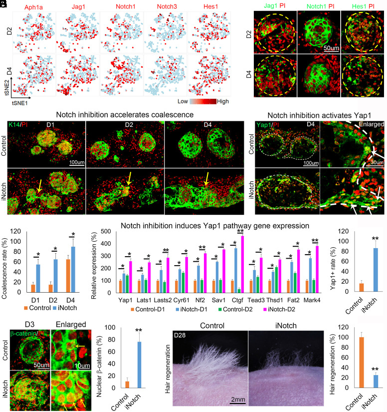
Stem cells in organoids self-organize into tissue patterns with unknown mechanisms. Here, we use skin organoids to analyze this process. Cell behavior videos show that the morphological transformation from multiple spheroidal units with morphogenesis competence (CMU) to planar skin is characterized by two abrupt cell motility-increasing events before calming down. The self-organizing processes are controlled by a morphogenetic module composed of molecular sensors, modulators, and executers. Increasing dermal stiffness provides the initial driving force (driver) which activates Yap1 (sensor) in epidermal cysts. Notch signaling (modulator 1) in epidermal cyst tunes the threshold of Yap1 activation. Activated Yap1 induces Wnts and MMPs (epidermal executers) in basal cells to facilitate cellular flows, allowing epidermal cells to protrude out from the CMU. Dermal cell-expressed Rock (dermal executer) generates a stiff force bridge between two CMU and accelerates tissue mixing via activating Laminin and β1-integrin. Thus, this self-organizing coalescence process is controlled by a mechano-chemical circuit. Beyond skin, self-organization in organoids may use similar mechano-chemical circuit structures.

Keywords: hair follicle development; organoids; self-organization; symmetry breaking; tissue fluidity.


Conflict of interest statement

The authors declare no competing interest.

Figures

Fig. 1.
Fig. 1.
Altered tissue fluidity during coalescence of multiple CMUs. (A) Schematic of examination from top view or side view of the skin organoid culture. (B) Setup of the two-photon microscopy for the time-lapse live imaging system. (C) Immunostaining for P-cadherin and schematic of self-organization process from polarization, vibration, protrusion, and coalescence. Yellow arrows point to the phenotypes. (D) Confocal images show the 3D rendering of cultured cells from K14H2BGFP transgenic mice. C (1-6) means CMUs (1-6). (E) Time-lapse live imaging shows the coalescence of two epidermal cysts (K14H2BGFP+) from D3 to D4. Red: Dermal cells infected with Lentivirus-RFP. (F, Left) Live imaging analysis of the cell motility during coalescence (https://www.pnas.​org/doi/10.1073/pnas.1700475114, from 78 to 96 h after culture. Modified from movies in ref. . (F, Right) Quantitative analyses of cell speed in different times; Eight cells were analyzed at different time periods including ①, ②, ③, and ④ depicted in the figure. The steady state is reached when the planar configuration is gradually achieved between days 5 and 9 in culture. The planar region is measured for phase 4. (G) Analysis of spatial velocity (speed and directionality) during the coalescence stage. The direction and length of black arrows indicate directionality and speed. (H) Cell shape in the CMUs that are about to start coalescence. P-cadherin immunostaining shows epithelial protrusions during coalescence. Yellow arrows show epithelial protrusions. (I) Cell shape analyses. Aspect ratio of the basal cells from CMUs at D2 and D4. On average, basal cells in D4 appear to be flatter, but they are not significantly different from those in D2. (J) Nuclear shape analyses. The aspect ratio of the cells in the leading state is higher than those in the middle and trailing regions during coalescence at D4. (K) Nucleus density in the coalescence zone, based on time-lapse videos at the coalescence stage. There is a decrease in cell numbers because cells disperse on the fused newplanar surface.
Fig. 2.
Fig. 2.
Bioinformatics analysis reveals key molecular changes during the transition process of coalescence. (A) RNA-seq analysis shows that 287 genes are up-regulated from D2 to D4. (B) RNA-seq analysis reveals differential gene expression of Mmps, Wnt ligands, and YAP-related pathway genes up-regulated at D4. (C) T-distributed stochastic neighbor embedding (t-SNE) plots show cell clustering of D2 and D4 samples. (D) T-SNE plots and immunostaining show mRNA (red) or protein expression of exemplary genes in basal cells (P-cadherin encoded by Cdh3), suprabasal cells (Klk7), and dermal fibroblasts (Collagen I) at D4. (E) ScRNA-seq analysis and immunostaining for Mmp9 at D2 and D4. (F) ScRNA-seq analysis and immunostaining for Lama5 at D2 and D4. *P < 0.05 and **P < 0.01; N ≥ 3. (G, Left) Quantification of Mmp9 expression at D4. (G, Middle) ScRNA-seq analysis of representative genes highly expressed in Mmp9+ vs. Mmp9- basal cells at D4. Right: Gene ontology (GO) analysis of signaling pathway in Mmp9+ cells.
Fig. 3.
Fig. 3.
Wnt induces Mmps expression to mediate the coalescence of CMUs. (A) ScRNA-seq analysis and immunostaining of Mmps expression in epidermal cells. (B) Immunostaining for K14 shows coalescence is inhibited at D4 and D7 after the inhibition of broad-spectrum MMPs. (C) Live imaging analysis of the decreased cell motility in the MMP inhibitor–treated group (iMMP, n≥ 8) compared with the control shown in Fig. 1F. (D) ScRNA-seq analysis and immunostaining of Wnts expression in epidermal cells. (E) qRT-PCR shows Mmps expression at D4 after addition of Wnt10a-P at D2. (F) Immunostaining and statistics reveal coalescence in Wnt10a-P-treated cultures. *P < 0.05. N ≥ 3.
Fig. 4.
Fig. 4.
Yap1 induces the coalescence of CMUs by activating the Wnt signaling pathway. (A) ScRNA-seq and immunostaining show exemplary YAP pathway gene expression. (B) Yap1 mRNA expression and gene-regulatory networks (GRNs) show that Yap pathway–activated genes are increased in basal epidermal cells from D2 to D4. (C) Immunostaining for YAP, β-catenin, MMP9, and Laminin expression at D4 using serial sections. (D) Quantitative RT-PCR shows the expression of Wnt pathway genes Wnts and β-catenin in Verteporfin-treated cells (treated at D2 and harvested at D4 and D6). (E) TSNE plot shows increased and overlapped expression of Yap1 and Wnt10a in basal cells from D2 to D4. KEGG analysis shows representative genes highly expressed in Yap1+ vs. Yap1- cells. (F) Immunostaining for β-catenin and MMP9 expression in Verteporfin-treated cells. (G) Time-lapse live imaging of K14H2BGFP epidermal cells after adding Verteporfin. (H) Immunostaining for Collagen XVII shows that inhibition of YAP1 leads to blocked coalescence at D4 and D6. Hair regeneration with transplantation of Verteporfin-treated cells (treated at D2, transplanted at D7, and observation at D21). **P < 0.01, ***P < 0.001, and # no significant difference. N ≥ 3.
Fig. 5.
Fig. 5.
Increasing dermal stiffness activates YAP1 in adjacent basal epidermal cells to initiate the symmetry-breaking process. (A) Measurement of stiffness within the culture using photonic crystal bio-force microscopy. (B) Quantification of nuclear YAP1 expression in Collagen I recombinant protein–treated cultures. (C) qRT-PCR reveals that expression of serval YAP1 pathway/target genes is significantly increased at D4 in Collagen I recombinant protein–treated cultures. (D) Quantification of epidermal protrusions in Collagen I recombinant protein–treated cultures. (E) Quantification of nuclear β-catenin+ cells in Collagen I recombinant protein–treated cultures. (F) Immunostaining for β-catenin and MMP9 expression in Collagen I recombinant protein–treated cultures. (G) Time-lapse live imaging shows that addition of Collagen I leads to accelerated coalescence at D4. (H) Hair regeneration with transplantation of Collagen I or Collagen IV recombinant protein–treated cultures (treated at D2, transplanted at D7, and observation at D21 after transplantation). *P < 0.05 and **P < 0.01. N ≥ 3.
Fig. 6.
Fig. 6.
Notch signaling in the basal layer can modulate YAP1 activation in epidermal cells. (A) T-SNE plots show representative Notch pathway gene expression in epidermal cells. (B) Immunostaining for Notch pathway gene expression in CMUs. (C) Inhibition of Notch (iNotch) results in accelerated coalescence. (D) Quantification of the coalescence rate after inhibition of Notch. (E) qRT-PCR reveals that mRNA expression of YAP1 pathway/target genes is significantly increased after Notch inhibition. (F) Immunostaining shows that Notch inhibition leads to increased Yap1 activation in basal epidermal cells. Yellow arrows show positive staining. (G) Quantification of nuclear YAP1+ cells in Notch-inhibited cultures. (H) Immunostaining shows that inhibition of Notch leads to increased β-catenin activation. (I) Hair regeneration after transplantation of Notch-inhibited cultures (treated at D0, transplanted at D7, and observation at D21 after transplantation). *P < 0.05 and **P < 0.01. N ≥ 3.
Fig. 7.
Fig. 7.
ROCK in dermal cells further promotes the mixing of CMU cells. (A) T-SNE plots and immunostaining show Rock1 and Rock2 expression (white arrow and pink schematic). (B) Time-lapse live imaging for K14GFP+ epidermal cells after ROCK inhibition (the white arrow indicates no coalescence). (C) Inhibition of Rock at D2 leads to blocked coalescence and giant CMUs formation. MMP14-induced epidermal protrusions cannot coalesce with Rock inhibition (the white arrow indicates the epidermal protrusions). (D) T-SNE plots show Lama4 and β1-integrin expression at D2 and D4. Immunostaining and statistics show that Rock activation results in relatively increased expression of Laminin and β1-integrin in dermal cells but accelerates basement membrane breakdown. **P < 0.01. N ≥ 3. (E) A summary model shows the driver, sensor, modulator, and executer for tissue fluidity during coalescence.

References

    1. Clevers H., Modeling development and disease with organoids. Cell 165, 1586–1597 (2016). - PubMed
    1. Kim J., Koo B. K., Knoblich J. A., Human organoids: Model systems for human biology and medicine. Nat. Rev. Mol. Cell Biol. 21, 571–584 (2020). - PMC - PubMed
    1. Bock C., et al. , The organoid cell atlas. Nat. Biotechnol. 39, 13–17 (2021). - PMC - PubMed
    1. Zhu M., Zernicka-Goetz M., Principles of self-organization of the mammalian embryo. Cell 183, 1467–1478 (2020). - PMC - PubMed
    1. Serra D., et al. , Self-organization and symmetry breaking in intestinal organoid development. Nature 569, 66–72 (2019). - PMC - PubMed

Publication types

Substances