. 2023 Sep 12;17(17):17451-17467.
doi: 10.1021/acsnano.3c05600. Epub 2023 Aug 29.

Exploiting Mass Spectrometry to Unlock the Mechanism of Nanoparticle-Induced Inflammasome Activation

Affiliations

Exploiting Mass Spectrometry to Unlock the Mechanism of Nanoparticle-Induced Inflammasome Activation

Govind Gupta et al. ACS Nano. .

Abstract

Nanoparticles (NPs) elicit sterile inflammation, but the underlying signaling pathways are poorly understood. Here, we report that human monocytes are particularly vulnerable to amorphous silica NPs, as evidenced by single-cell-based analysis of peripheral blood mononuclear cells using cytometry by time-of-flight (CyToF), while silane modification of the NPs mitigated their toxicity. Using human THP-1 cells as a model, we observed cellular internalization of silica NPs by nanoscale secondary ion mass spectrometry (nanoSIMS) and this was confirmed by transmission electron microscopy. Lipid droplet accumulation was also noted in the exposed cells. Furthermore, time-of-flight secondary ion mass spectrometry (ToF-SIMS) revealed specific changes in plasma membrane lipids, including phosphatidylcholine (PC) in silica NP-exposed cells, and subsequent studies suggested that lysophosphatidylcholine (LPC) acts as a cell autonomous signal for inflammasome activation in the absence of priming with a microbial ligand. Moreover, we found that silica NPs elicited NLRP3 inflammasome activation in monocytes, whereas cell death transpired through a non-apoptotic, lipid peroxidation-dependent mechanism. Together, these data further our understanding of the mechanism of sterile inflammation.

Keywords: cell death; inflammasome; mass spectrometry; monocyte; silica nanoparticles.


Conflict of interest statement

The authors declare the following competing financial interest(s): M.P. is a former employee of Nouryon PPC, a company producing colloidal silica. Nouryon PPC had no input on the paper. The other authors declare no conflicts of interest.

Figures
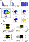
Figure 1
Figure 1
Single-cell mass cytometry of silica NP-exposed PBMCs. (a) Cell viability analysis of primary human monocytes after 24 h exposure to uncoated/bare, Al-doped, and silane-modified silica NPs. Data shown are mean values ± SD of experiments performed using cells from three independent donors. **p < 0.01; ***p < 0.001. (b) viSNE analysis depicting the single-cell characterization of PBMCs. For gating strategies for immune cell subpopulations, refer to Figure S5. (c) Single-cell analysis of cell viability by CyTOF. PBMCs were treated with bare or ethoxysilane (ES)-modified silica NPs at 0.1 μg/mL for 24 h. The viSNE plots show the different immune cell subpopulations for treated or untreated samples. LPS was used as a positive control. (d–g) Heat maps and histograms of rhodium mean marker expression ratios for gated T-cell subpopulations (d), monocyte subpopulations (e), DC and NK cell populations (f), and B cell subpopulations (g).
Figure 2
Figure 2
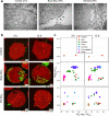
Cellular interaction and uptake of small silica NPs. (a) TEM images showing cell surface interactions of bare versus silane-modified silica NPs at 2 h. Arrows point to NPs on the cell membrane or outside the cells. For results on other silica NPs of different primary particle sizes, refer to Figure S8. (b) Label-free nanoSIMS analysis of THP-1 cells exposed to bare and silane-modified silica NPs for 2 and 12 h. (c) Quantification of relative abundance of silica NP distribution in cells at 2 and 12 h, based on nanoSIMS.
Figure 3
Figure 3
Silica NPs trigger cytoplasmic lipid droplet formation. (a) TEM images showing ultrastructural changes in THP-1 cells exposed for 12 h to bare silica NPs versus control cells. Cytoplasmic vacuolization (upper panel) and lipid droplet accumulation (arrows, lower panel) were noted. (b, c) Lipid droplet content quantified using flow cytometry after labeling the cells with BODIPY 493/503. The cells were exposed to silica NPs in the presence or absence of Trolox. For further results using Nile Red staining, see Figure S10.
Figure 4
Figure 4
Silica NP-induced membrane lipid changes analyzed by ToF-SIMS. (a) ToF-SIMS mass spectra recorded in positive ion (m/z 550 to m/z 850) indicating that phosphatidylcholine (PC) and its fragments were decreased after exposure for 6 h to small (12 nm) bare SiO2 NPs (blue) with respect to control (untreated cells) (gray). (b, c) Heat maps showing normalized intensity values of sn-1 and sn-2 acyl chain fatty acids detected in negative ion mode at 2 and 6 h of exposure, respectively. THP-1 cells were exposed to bare and ethoxysilane (ES)-modified silica NPs in the presence and absence of Trolox, as indicated. Refer to Figure 5 for quantification of the data obtained at 6 h.
Figure 5
Figure 5
ToF-SIMS reveals changes in fatty acids in the plasma membrane of silica NP-exposed cells. (a) ToF-SIMS mass spectra recorded in negative ion mode (m/z 150 to m/z 350) indicating a change in various fatty acids at 6 h after exposure to small, bare SiO2 NPs (blue) with respect to control (gray). (b, c) Most significantly altered unsaturated and saturated fatty acids, respectively, following exposure of THP-1 cells to bare versus silane-modified (ES) silica NPs for 6 h, in the presence or absence of Trolox. The results in panels b and c are shown as mean values ± SD (n = 4). *p < 0.05; **p < 0.01; ****p < 0.0001.
Figure 6
Figure 6
Silica NPs trigger NLRP3-dependent IL-1β secretion. IL-1β release triggered by bare and Al-doped silica NPs is blocked by the cathepsin B inhibitor, CA074Me (a), the pan-caspase inhibitor, zVAD-fmk (b), and MCC950, a selective inhibitor of the NLRP3 inflammasome (c). For results on other silica NPs of varying sizes as well as the benchmark NPs, refer to Figure S16. Note that the cells were nonprimed. For results using LPS-primed cells, refer to Figure S17. The role of the NLRP3 inflammasome (in the absence of LPS priming) was further confirmed by using wild-type (Null-1) and NLRP3-deficient THP-1 cells (d). For results using caspase-1-deficient cells, refer to Figure S18. (e) Lipid antioxidant Trolox significantly reduced IL-1β release triggered by bare silica NPs. (f) Cell death was also significantly reduced by Trolox, while cell death unaffected by cathepsin B or caspase inhibitors (see Figure S12). Data are shown as mean values ± SD (n = 3). ***p < 0.001; ###p < 0.001; ****p < 0.0001; #p < 0.05; ####p < 0.0001.
Figure 7
Figure 7
Pro-inflammatory responses of silica NPs are iPLA2-dependent. (a) Bare silica NPs triggered an elevation in cellular LPC content in THP-1 cells, which was prevented by BEL, a selective iPLA2-VIA inhibitor. Cells were exposed for 6 h to bare silica NPs (2.5 μg/mL) with or without BEL (5 μM). (b) Silica NP-triggered IL-1β release in nondifferentiated (monocyte-like) THP-1 cells was effectively blocked by BEL. (c) Silica NP-triggered IL-1β release in primary human CD14+ monocytes was also reduced by BEL. (d) Western blot to confirm PLA2G6 silencing in THP-1 cells using specific siRNAs. GAPDH was used as a loading control. (e) Silencing of PLA2G6 significantly reduced IL-1β production as determined by ELISA. (f) Silica NPs triggered inflammasome assembly at the MTOC, as evidenced by the colocalization of ASC and the centrosomal marker, γ-tubulin (GTU). Samples obtained after 6 h of exposure were visualized by confocal microscopy. For additional results on the impact of BEL, refer to Figure S20. Data shown as mean values ± SD (n = 3). **p < 0.01; ****p < 0.0001; ##p < 0.01; ####p < 0.0001.
Figure 8
Figure 8
Understanding sterile inflammation. Schematic of the classical two-signal model of inflammasome activation (a) versus the present model (b). The NLRP3 inflammasome consisting of NLRP3, ASC, and pro-caspase-1 serves as a platform for the activation of caspase-1, leading to the proteolysis of pro-IL-1β and the release of IL-1β. Numerous studies have shown that particulate matter (PM) such as alum and crystalline silica (quartz) as well as high aspect ratio (nano)materials (HARNs), e.g., asbestos and carbon nanotubes, are able to trigger the activation of the NLRP3 inflammasome (panel a). This usually occurs through the internalization of the offending agent into phagosomes that eventually fuse with lysosomes (the phago-lysosomal system is depicted here as an empty circle), leading to the generation of reactive oxygen species (ROS) and the release of lysosomal cathepsin B. Macrophages are usually primed with a Toll-like receptor (TLR) agonist such as LPS (lipopolysaccharide). In contrast, we show that uncoated amorphous silica NPs are capable of triggering NLRP3-dependent IL-1β release in the absence of LPS priming (panel b) and we provide evidence that mitochondrial ROS production may contribute to the effects of silica NPs on membrane lipids (mitochondria are depicted here as a small empty circle). We posit that lipid peroxidation, leading to phospholipase (iPLA2) activation and the generation of lysophosphatidylcholine (LPC), provides a cell autonomous “priming” signal.

References

    1. Broz P.; Dixit V. M. Inflammasomes: Mechanism of Assembly, Regulation and Signalling. Nat. Rev. Immunol. 2016, 16, 407–420. 10.1038/nri.2016.58. - DOI - PubMed
    1. Sun B.; Wang X.; Ji Z.; Li R.; Xia T. NLRP3 Inflammasome Activation Induced by Engineered Nanomaterials. Small 2013, 9, 1595–1607. 10.1002/smll.201201962. - DOI - PMC - PubMed
    1. Dostert C.; Pétrilli V.; Van Bruggen R.; Steele C.; Mossman B. T.; Tschopp J. Innate Immune Activation Through Nalp3 Inflammasome Sensing of Asbestos and Silica. Science 2008, 320, 674–677. 10.1126/science.1156995. - DOI - PMC - PubMed
    1. Cassel S. L.; Eisenbarth S. C.; Iyer S. S.; Sadler J. J.; Colegio O. R.; Tephly L. A.; Carter A. B.; Rothman P. B.; Flavell R. A.; Sutterwala F. S. The Nalp3 Inflammasome is Essential for the Development of Silicosis. Proc. Natl. Acad. Sci. U. S. A. 2008, 105, 9035–9040. 10.1073/pnas.0803933105. - DOI - PMC - PubMed
    1. Hornung V.; Bauernfeind F.; Halle A.; Samstad E. O.; Kono H.; Rock K. L.; Fitzgerald K. A.; Latz E. Silica Crystals and Aluminum Salts Activate the NALP3 Inflammasome Through Phagosomal Destabilization. Nat. Immunol. 2008, 9, 847–856. 10.1038/ni.1631. - DOI - PMC - PubMed

Publication types