. 2023 Sep 13;23(17):8115-8125.
doi: 10.1021/acs.nanolett.3c02193. Epub 2023 Aug 29.

Accurate and Convenient Lung Cancer Diagnosis through Detection of Extracellular Vesicle Membrane Proteins via Förster Resonance Energy Transfer

Affiliations

Accurate and Convenient Lung Cancer Diagnosis through Detection of Extracellular Vesicle Membrane Proteins via Förster Resonance Energy Transfer

Shuting Xiao et al. Nano Lett. .

Abstract

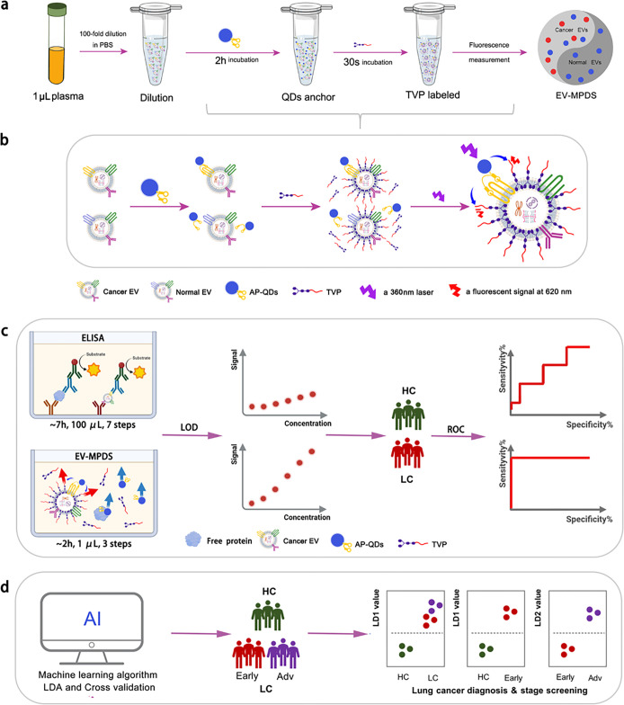
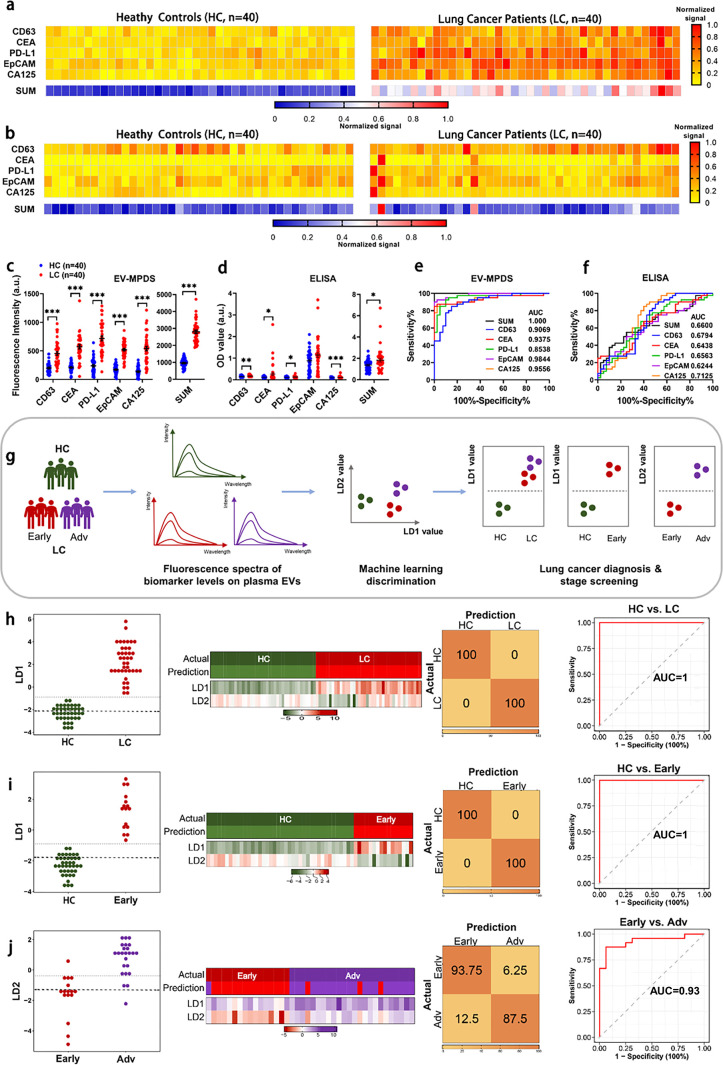
Tumor-derived extracellular vesicles (EVs) are promising to monitor early stage cancer. Unfortunately, isolating and analyzing EVs from a patient's liquid biopsy are challenging. For this, we devised an EV membrane proteins detection system (EV-MPDS) based on Förster resonance energy transfer (FRET) signals between aptamer quantum dots and AIEgen dye, which eliminated the EV extraction and purification to conveniently diagnose lung cancer. In a cohort of 80 clinical samples, this system showed enhanced accuracy (100% versus 65%) and sensitivity (100% versus 55%) in cancer diagnosis as compared to the ELISA detection method. Improved accuracy of early screening (from 96.4% to 100%) was achieved by comprehensively profiling five biomarkers using a machine learning analysis system. FRET-based tumor EV-MPDS is thus an isolation-free, low-volume (1 μL), and highly accurate approach, providing the potential to aid lung cancer diagnosis and early screening.

Keywords: Förster resonance energy transfer; aptamer; extracellular vesicles; lung cancer diagnosis; membrane proteins.


Conflict of interest statement

The authors declare no competing financial interest.

Figures

Figure 1
Figure 1
Schematic illustration of EV-MPDS. (a) Procedures of EV-MPDS work. (b) Rationale of EV-MPDS via FRET, with aptamer QDs serving as the donor molecule and TVP as the acceptor molecule. (c) EV-MPDS showed lower detection limitation and higher ability of lung cancer diagnosis compared with ELISA. (d) EVMPDS assisted in lung cancer diagnosis, early detection, and staging based on the AI classification.
Figure 2
Figure 2
EV-MPDS confirmation. (a) TEM images of A549 EV and CD63 aptamer-QDs labeled A549 EVs. (b) PL spectra of CD63 aptamer QDs at 360 nm laser excitation. (c) Absorption spectra of TVP in the aqueous solutions. (d) PL spectra of TVP-labeled EVs at 465 nm laser excitation. (e) Fluorescence spectra of the QDs, TVPs, and QDs with TVP-labeled EVs under 360 nm laser excitation. (f) Comparison of 620 nm fluorescence intensity of different TVP-labeled samples under excitation at 360 nm. (g) Fluorescence intensity comparison of EVs mixed with BSA at the different concentrations co-labeled with CD63-QDs and TVP. (h) CLSM images of A549 cells labeled with QDs only, TVP only, or both compounds sequentially. (QDs channel: Ex = 405 nm, Em = 450–525 nm; E-FRET channel: Ex = 405 nm, Em = 550–700 nm; TVP channel: Ex = 488 nm, Em = 550–700 nm; scale bar = 10 μm).
Figure 3
Figure 3
Performance of EV-MPDS was evaluated. (a, b) TEM images (a) and nanoparticle tracking analysis (b) were conducted to visualize and analyze the EVs extracted from A549 cell supernatant. (c, d) Fluorescence spectra (c) and the relationship between fluorescence intensity and EV concentration (d) were measured for EVs labeled with QDs only at different concentrations. (e, f) The same analysis was performed for EVs detected by EV-MPDS. (g) ELISA analysis was used to determine the relationship between fluorescence intensity and EV concentration. (h–m) PD-L1 was detected on EVs extracted from equal volumes of A549, H1299, H23, and Beas-2b cell supernatant using EV-MPDS, ELISA, and WB. (h) Fluorescence spectra, (i) CLSM images were obtained for the four types of EVs, and (j) the fluorescence intensity analysis in EV-MPDS detection. The PD-L1 expression levels in different samples were analyzed by (k) ELISA and (l) Western blot with (m) representative Western blot images.
Figure 4
Figure 4
Detection and classification verification of EV-MPDS in unextracted mixture samples were performed. (a) The process involved preparing and detecting the cell supernatant concentrate before extraction and extractive EVs. (b–e) PD-L1 liposomes were used to mimic PD-L1 on EVs. (b, c) WB analysis, (d) ELISA analysis, and (e) EV-MPDS were conducted to examine liposomes, PD-L1 liposomes, PD-L1, and a mixture of PD-L1 and PD-L1 liposomes. (f, g) PD-L1 abundance was evaluated in the cell supernatant and EVs extracted from the cell supernatant using ELISA (f) and EV-MPDS (g), respectively. (h, i) Four cancer biomarkers (CEA, PD-L1, EpCAM, and CA125) on EVs to distinguish lung cancer cell lines from normal cell line by WB analysis. (h) Representative blot images and (i) a heat map were generated to show the expression of these biomarkers in cell and EV samples. (j–m) Scatter plots were used to verify the expression distribution of biomarkers (CEA, PD-L1, EpCAM, and CA125) in the cell supernatant and EVs extracted from the cell supernatant, as detected by ELISA and EV-MPDS, respectively (# shows the significant differences compared to the Beas 2b group). The study included A549, H1299, and H23 as cancer cell lines and Beas-2b as the normal cell line.
Figure 5
Figure 5
Diagnosis of lung cancer involves detecting circulating EVs using EV-MPDS and AI classification. A study examined EV samples from 40 lung cancer patients and 40 healthy controls, assessing five key lung cancer protein markers (CD63, CEA, PD-L1, EpCAM, and CA125). (a, b) Heat maps were generated to show cancer biomarker protein profiles using (a) EV-MPDS and (b) ELISA methods. (c, d) Scatter plots of the five EV biomarkers were created for plasma samples analyzed by (c) EV-MPDS and (d) ELISA. (e, f) ROC curve analysis evaluated the diagnostic power of the individual biomarkers and the SUM signature using (e) EV-MPDS and (f) ELISA. (g) Flowchart of signals obtained by EV-MPDS followed by a weighted ensemble classification system. (h–j) LDA-based analysis of EV biomarkers was used for lung cancer detection, providing LD1 or LD2 values for control and lung cancer group samples. Heat maps, confusion matrices, and ROC curves were generated for different comparisons, including (h) control vs lung cancer group, (i) control vs early stage lung cancer group, and (j) early stage vs advanced-stage lung cancer group.

Similar articles

Cited by

References

    1. Sung H.; Ferlay J.; Siegel R. L.; Laversanne M.; Soerjomataram I.; Jemal A.; Bray F. Global Cancer Statistics 2020: GLOBOCAN Estimates of Incidence and Mortality Worldwide for 36 Cancers in 185 Countries. CA Cancer J. Clin. 2021, 71 (3), 209–249. 10.3322/caac.21660. - DOI - PubMed
    1. Pastorino U.; Silva M.; Sestini S.; Sabia F.; Boeri M.; Cantarutti A.; Sverzellati N.; Sozzi G.; Corrao G.; Marchianò A. Prolonged lung cancer screening reduced 10-year mortality in the MILD trial: new confirmation of lung cancer screening efficacy. Ann. Oncol 2019, 30 (7), 1162–1169. 10.1093/annonc/mdz117. - DOI - PMC - PubMed
    1. Crosby D.; Bhatia S.; Brindle K. M.; Coussens L. M.; Dive C.; Emberton M.; Esener S.; Fitzgerald R. C.; Gambhir S. S.; Kuhn P.; Rebbeck T. R.; Balasubramanian S. Early detection of cancer. Science 2022, 375 (6586), eaay9040.10.1126/science.aay9040. - DOI - PubMed
    1. Xue Y.; Feng X.; Fan X.; Zhu G.; McLaughlan J.; Zhang W.; Chen X. Extracellular Vesicles for the Diagnosis of Cancers. Small Structures 2022, 3 (1), 2100096.10.1002/sstr.202100096. - DOI
    1. Kalluri R.; LeBleu V. S. The biology, function, and biomedical applications of exosomes. Science 2020, 367 (6478), eaau6977.10.1126/science.aau6977. - DOI - PMC - PubMed

Publication types

Substances