. 2023 Oct 10;56(10):2311-2324.e6.
doi: 10.1016/j.immuni.2023.08.001. Epub 2023 Aug 28.

Mechanotransduction via endothelial adhesion molecule CD31 initiates transmigration and reveals a role for VEGFR2 in diapedesis

Affiliations

Mechanotransduction via endothelial adhesion molecule CD31 initiates transmigration and reveals a role for VEGFR2 in diapedesis

Tao Fu et al. Immunity. .

Abstract

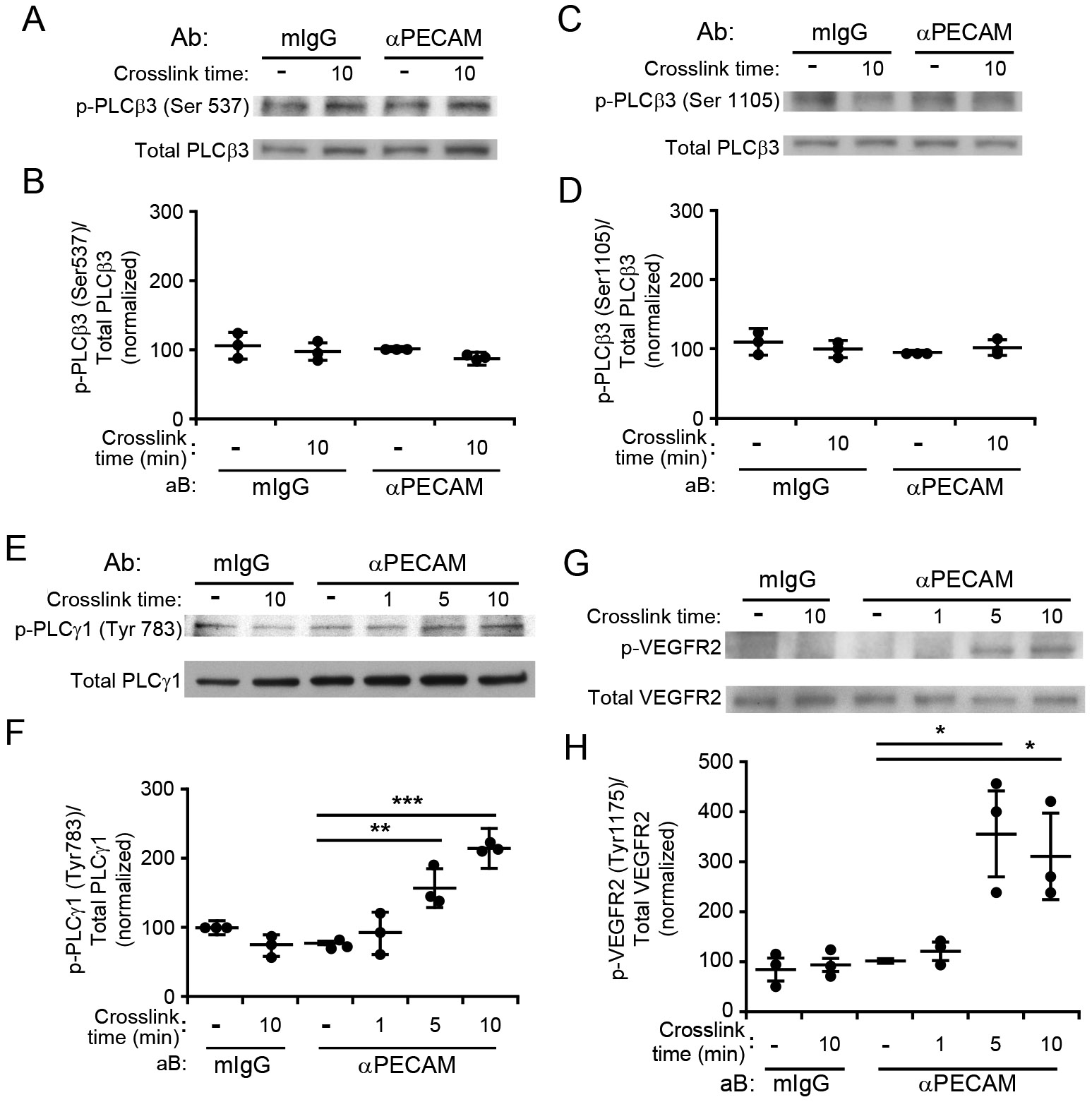
Engagement of platelet endothelial cell adhesion molecule 1 (PECAM, PECAM-1, CD31) on the leukocyte pseudopod with PECAM at the endothelial cell border initiates transendothelial migration (TEM, diapedesis). We show, using fluorescence lifetime imaging microscopy (FLIM), that physical traction on endothelial PECAM during TEM initiated the endothelial signaling pathway. In this role, endothelial PECAM acted as part of a mechanotransduction complex with VE-cadherin and vascular endothelial growth factor receptor 2 (VEGFR2), and this predicted that VEGFR2 was required for efficient TEM. We show that TEM required both VEGFR2 and the ability of its Y1175 to be phosphorylated, but not VEGF or VEGFR2 endogenous kinase activity. Using inducible endothelial-specific VEGFR2-deficient mice, we show in three mouse models of inflammation that the absence of endothelial VEGFR2 significantly (by ≥75%) reduced neutrophil extravasation by selectively blocking diapedesis. These findings provide a more complete understanding of the process of transmigration and identify several potential anti-inflammatory targets.

Keywords: PECAM; VEGFR2; endothelial cell; inflammation; inflammatory signaling; leukocyte; mechanotransduction; monocyte; neutrophil; transmigration.


Conflict of interest statement

Declaration of interests The authors declare no competing interests.

Figures

Figure 1.
Figure 1.. Cross-linking endothelial cell PECAM overcomes a PECAM antibody block in a calcium-dependent manner and leads to PLC-dependent TEM.
(A) HUVEC transfected to express FLAG-tagged TRPC6 were incubated with anti-FLAG, anti-PECAM, or both (see Methods) then incubated with appropriate secondary antibodies and subjected to the PLA procedure. Negative controls incubated with either antibody and control IgG gave no signal (small upper panels). However, strong junctional distribution (arrows) of fluorescence was seen in the monolayers that received both antibodies, indicating that PECAM and TRPC6 closely co-localize at endothelial cell borders. Scale bar represents 50 μm. (B) Scheme showing the methodology of the antibody blockade and crosslinking TEM assays. (Bi) Interaction of leukocyte PECAM with EC PECAM activates TRPC6 to allow ↑[Ca2+]i and promote TEM. (Bii) Blocking anti-PECAM antibodies prevent interaction with leukocyte PECAM. No PECAM signal is generated, there is no ↑[Ca2+]i and no TEM. (Biii) When the same PECAM antibodies are cross-linked, it mimics the engagement provided by leukocyte PECAM activating the endothelial PECAM signal, activating TRPC6, and promoting TEM. (Biv) When the same experiment is performed in EC in which calcium concentration is buffered by BAPTA, the critical ↑[Ca2+]i is not achieved and there is no TEM. Neutrophils (C) and monocytes (D) were added to HUVEC monolayers pretreated for 40 min with either BAPTA or vehicle and/or PECAM blocking antibodies or non-blocking anti-VE-cadherin isotype control. After 10 min (for neutrophils) or 40 min (for monocytes) goat anti-mouse F(ab’)2 was added to cross-link PECAM. Data shown represent five and four replicates for neutrophils and monocytes, respectively, with at least 300 leukocytes counted per replicate. Data are the average and standard deviation of at least three replicates per sample (typically four or five). Dose response analysis showing the effect of the broad-spectrum PLC inhibitor (U73122) on the TEM (E and G) and adhesion (F and H) of neutrophils (E and F) and monocytes (G and H). HUVEC were pretreated with either U71322 or vehicle (DMSO), or blocking anti-PECAM mAb as indicated. Quantitative data are representative of the mean and standard deviation of three experiments where at least 300 hundred leukocytes were scored per condition. Data represent the average and standard deviation of three experiments (PMN) and two experiments (monocytes). (I) Eluate control experiment (see Methods--TEM) to show that negligible U73122 elutes from the HUVEC during the experiment and does not affect the leukocytes in this TEM assay. Dots represent means of individual experiments unless otherwise indicated. Bars are the mean and standard deviation. Asterisks ** and *** denote p-values <0.01 and <0.001 respectively.
Figure 2.
Figure 2.. PECAM cross-linking induces phosphorylation of PLCγ and VEGFR2.
HUVEC monolayers were incubated with either anti-PECAM antibodies or control non-specific mouse IgG (mIgG) for 1 h and then washed and treated with secondary cross-linking antibody for the indicated time to stimulate PECAM signaling. Monolayers were then lysed and their proteins resolved using SDS-PAGE and western blotting. The phosphorylation of PLCβ3 at serines 537 (A) and 1105 (C) PLCγ1 tyrosine 783 (E), and VEGFR2 tyrosine 1175 (G) were probed using specific antibodies. Blots were then stripped and reprobed to determine total protein expression. Band intensities were quantified using densitometry and normalized to the total amount of the indicated protein (B, D, F, H). Data shown were normalized to the control (mIgG, no cross-linking) samples. Dots represent means of individual experiments unless otherwise indicated. Bars are the mean and standard deviation. Asterisks *, **, and *** denote p-values < 0.05, <0.01 and <0.001 respectively.
Figure 3.
Figure 3.. PECAM tension sensing is required for TEM.
(A) Schematic of PECAM tension sensing and control constructs used in this project. (B) HUVEC monolayers were transfected with PECAM shRNA followed by PECAM tension sensor (TS) and non-sensing (Δ15,16) rescue constructs as indicated. (B) Western blot analysis of transfected constructs confirms efficient protein depletion and reexpression. Samples were probed with GAPDH as a loading control. (C) Monolayers prepared as described in (B) were subjected to shear flow at 11.5 dynes/cm2 for 48 h as described in the Materials and Methods. They were then fixed, stained for VE-cadherin (blue) and actin (red), and visualized using confocal microscopy. Arrow indicates direction of flow; scale bar is 100 μm. (D) Quantitation of the inverse aspect ratio in C, where 1.0 would be a cell with equal length and width. Data are the mean and standard deviation of at least 50 cells from three separate experiments. (E) HUVEC monolayers prepared as described in B were incubated with monocytes for one hour to examine their ability to support TEM. Data shown represent the mean and standard deviation of three separate experiments with at least 300 monocytes scored per experiment. (F,G) HUVEC monolayers prepared as described above were visualized by fluorescence lifetime imaging microscopy (FLIM) measurements during neutrophil TEM. Only monolayers expressing the tension-sensing PECAM construct showed an increase in fluorescence lifetime of TFP, indicating stretching of the PECAM molecule that corresponded to the act of TEM (F) Dots represent individual PMN/EC interactions. At least 11 were observed for each condition. (G) Image of PECAM tension sensor around a neutrophil (red; yellow asterisk) engaged in TEM. The dashed yellow circle denotes the corresponding region of interest that was measured for FLIM. Scale bar = 10 μm. Dots represent means of individual experiments unless otherwise indicated. Bars are mean ± standard deviation. Asterisks ** and *** denote p-values <0.01 and <0.001 respectively.
Figure 4.
Figure 4.. Efficient TEM requires the endogenous transmembrane domain of VE-cadherin.
HUVEC were incubated with adenovirus expressing shRNA against VE-cadherin and then rescue constructs that expressed either wild-type VE-cadherin (WT-TM) or VE-cadherin with its transmembrane domain replaced with that of N-cadherin (N-cad-TM). (A) Representative (of 3 independent experiments) Western blot showing efficient depletion of endogenous VE-cadherin and appropriate re-expression of TM-domain constructs. (B) Quantification of TEM assays using endothelial monolayers prepared as in (A). shRNA depletion of endogenous VE-cadherin significantly impairs TEM. Re-expression of wild-type VE-cadherin restored TEM to baseline, whereas re-expression of VE-cadherin whose transmembrane domain was replaced with that of N-cadherin did not. Dots represent means of individual experiments unless otherwise indicated. Bars are mean and standard deviation from three independent experiments. Asterisks ** denote p-value <0.01.
Figure 5.
Figure 5.. VEGFR2 is required for targeted recycling and TEM in a ligand-independent manner.
(A) Depletion of VEGFR2 in HUVEC using shRNA delivered by adenovirus and reconstitution of those HUVEC with wild-type VEGFR2 (WT) or VEGFR2 with tyrosine 1175 mutated to phenylalanine (Y1175F). (B) Neutrophils (PMN) subjected to transmigration assay across HUVEC monolayers expressing VEGFR2 as in panel (A). TEM is significantly diminished in the VEGFR2 depleted cells, but rescued completely when WT VEGFR2 is restored. Re-expressing VEGFR2 with the Y1175F mutation does not rescue TEM. (C) Running the TEM assay in the presence or absence of anti-VEGF antibody did not affect transmigration of PMN. (D) HUVEC serum-starved overnight then pre-incubated with 10 μg/ml of bevacizumab, a blocking anti-VEGF antibody or 10 μM SU1498 for 20 minutes prior to addition of VEGF (20 ng/ml). Incubation of HUVEC for 5 min. Monolayers were lysed and subjected to Western Blot for p-1175 VEGFR2, then stripped and probed for total VEGFR2. (E) TEM assays of monocytes were carried out in the presence of SU1498 at the concentrations indicated, or hec7 (blocking anti-PECAM mAb) at 20 μg/ml for 55 minutes. After 55 minutes, Hyp9 or YODA1 were added to the corresponding wells to a final concentration of 10 μM in the continued presence of hec7. The TEM assay was stopped at 60 min. (F) Control HUVEC monolayers or those with VEGFR2 depleted and replaced with the indicated rescue constructs (K-D = kinase dead) underwent PECAM cross-linking and blotting for phosphorylation of Y1175. Results are representative of 3 independent experiments. (G) HUVEC monolayers with VEGFR2 depleted and rescued with control virus, wild-type VEGFR2, or the indicated mutant VEGFR2 constructs were used in the TEM assay. (H) Control experiment to show that kinase dead (K-D) VEGFR2 mutant is not phosphorylated when exposed to VEGF. (I) TEM of neutrophils across HUVEC pretreated with DMSO (Control), or 5 μM PP2 or PP3 for 10 min. Eluate controls were performed as in Fig. 1I, showing that no detectable reagent eluted from the monolayers to directly affect the leukocytes. (J) HUVEC were loaded with Fluo4 and imaged live using a water-immersion lens for several minutes before and after addition of DMSO (carrier), YODA1 (10 μM), and histamine (10 μM) Still images from the live recording (Please see Supplemental Videos 1-3) are shown. Scale bar is 100 μm. (K, L) Targeted recycling assay during transmigration of PMN across control, VEGFR2 depleted and depleted→reconstituted endothelial monolayers. (K) During normal TEM (Control), the LBRC moves to surround the PMN as it transmigrates, while VE-cadherin forms a gap (arrowhead). When VEGFR2 is depleted (VEGFR2 KD), the LBRC does not move to the surface to surround the PMN and VE-cadherin remains visible under the neutrophil (arrowhead). The result is no TEM. Scale bar is 20 μm. (L) Quantification of the percent of PMN with enriched LBRC on the corresponding monolayers. Because this assay captures a snapshot in time early in the transmigration of the PMN population, most PMN have not transmigrated yet. However, the ratios of LBRC enrichment are proportional to the final TEM in this assay (panel B). Asterisks * and *** denote p-values of <0.05 and <0.001 respectively.
Figure 6.
Figure 6.. Endothelial-specific inducible VEGFR2 deficient mice have defective neutrophil extravasation.
(A) Inducible endothelial-specific Cre mice (Cdh5(PAC)-CreERT2 were mated with Flk-1fl/fl mice and backcrossed until homozygous for Flk-1fl/fl. Mice were treated with tamoxifen (+TAM) in corn oil to induce deficiency of VEGFR2 selectively in endothelial cells (iECKO) or corn oil alone for 5 days, rested for at least two weeks, and then endothelial cells isolated from lungs. Lysates were probed by Western Blot for VEGFR2. (B) Mice with inducible endothelial-specific deficiency of VEGFR2 (iECKO) or littermates that received corn oil only were studied in the croton oil dermatitis model. Five hours after application of croton oil to the mouse ear skin many PMN (S100A9, green) were seen in the interstitial tissue outside of vessels (PECAM, red) and their associated basement membrane (collagen IV, blue). An equal number of PMN are recruited to the ears of inducible endothelial-specific VEGFR2 deficient mouse, but the vast majority remain within the postcapillary venules, apparently attached to the endothelium. Scale bar is 100 μm. (C) Quantification of three independent experiments. PMN extravasation is reduced from 80% to 20%. Asterisk denotes p-value <0.01. (D) Inducible endothelial-specific VEGFR2 deficient mice bred as in (A) were fed Tamoxifen Chow or normal chow for at least 20 days, then PBS (Control) or 0.1N HCl was instilled into their lungs via the trachea. (See Methods.) Twenty four hours later, mice were euthanized and bronchoalveolar lavage (BAL) fluid collected from each mouse. The lungs were then harvested, extracted, and probed for VEGFR2 by Western blot. Graph (D) shows quantification of blots for 8 mice per condition. The number of neutrophils (E) and monocytes (F) was quantified from the BAL fluid. The dots represent data from individual mice in an experiment representative of three independent experiments. Bars represent mean ± standard deviation. Asterisks ** and *** denote p-values of <0.01 and <0.001 respectively.
Figure 7.
Figure 7.. Endothelial-specific inducible VEGFR2 deficient mice have defective transendothelial migration.
(A) EC-specific VEGFR2-deficient mice (iECKO) were produced as for the experiments in Fig. 6. Male mice were studied by intravital microscopy of the cremaster muscle circulation following intrascrotal injection of murine IL-1β. Video recordings were made using a spinning disc confocal microscope as described . Shown are representative frames from Supplemental Videos 4 and 5. Circulating PMN express LysM-eGFP (green); endothelial cell borders are stained by a non-blocking mAb against mouse PECAM (red). White arrowheads point to extravasating PMN. (B – D) Quantification of rolling (B), adhesion (C), and TEM (D) from at least three independent experiments. Dots represent means of individual experiments unless otherwise indicated. Bars are the mean ± standard deviation is shown. Only the TEM step was significantly decreased in the endothelial specific inducible VEGFR2-deficient mice. Asterisk * denotes p-value <0.05, scale bar = 50 μm.

References

    1. Huang AJ, Manning JE, Bandak TM, Ratau MC, Hanser KR, and Silverstein SC (1993). Endothelial cell cytosolic free calcium regulates neutrophil migration across monolayers of endothelial cells. J.Cell Biol 120, 1371–1380. - PMC - PubMed
    1. Muller WA, Weigl SA, Deng X, and Phillips DM (1993). PECAM-1 is required for transendothelial migration of leukocytes. J Exp Med 178, 449–460. 10.1084/jem.178.2.449. - DOI - PMC - PubMed
    1. Weber EW, Han F, Tauseef M, Birnbaumer L, Mehta D, and Muller WA (2015). TRPC6 is the endothelial calcium channel that regulates leukocyte transendothelial migration during the inflammatory response. J Exp Med 212, 1883–1899. 10.1084/jem.20150353. - DOI - PMC - PubMed
    1. Dalal PJ, Sullivan DP, Weber EW, Sacks DB, Gunzer M, Grumbach IM, Heller Brown J, and Muller WA (2021). Spatiotemporal restriction of endothelial cell calcium signaling is required during leukocyte transmigration. J Exp Med 218, e20192378. 10.1084/jem.20192378. - DOI - PMC - PubMed
    1. Tzima E, Irani-Tehrani M, Kiosses WB, Dejana E, Schultz DA, Engelhardt B, Cao G, DeLisser H, and Schwartz MA (2005). A mechanosensory complex that mediates the endothelial cell response to fluid shear stress. Nature 437, 426–431. - PubMed

Publication types

MeSH terms

Substances

LinkOut - more resources