

Use of mesenchymal stem cells for tendon healing in veterinary and human medicine: getting to the "core" of the problem through a one health approach
- PMID: 37643722
- PMCID: PMC11027114
- DOI: 10.2460/javma.23.07.0388
Use of mesenchymal stem cells for tendon healing in veterinary and human medicine: getting to the "core" of the problem through a one health approach
Abstract
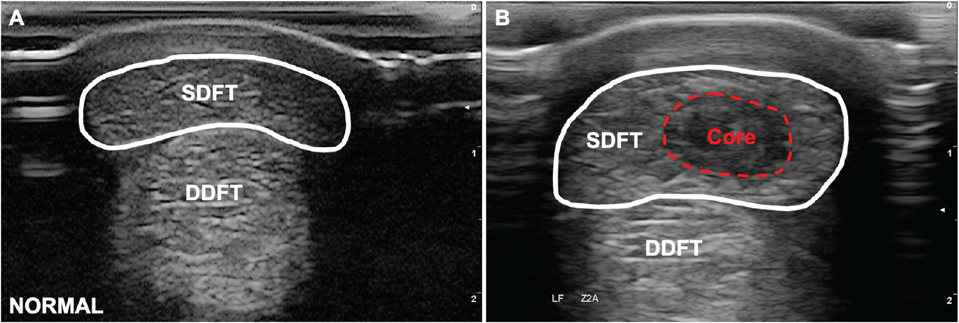
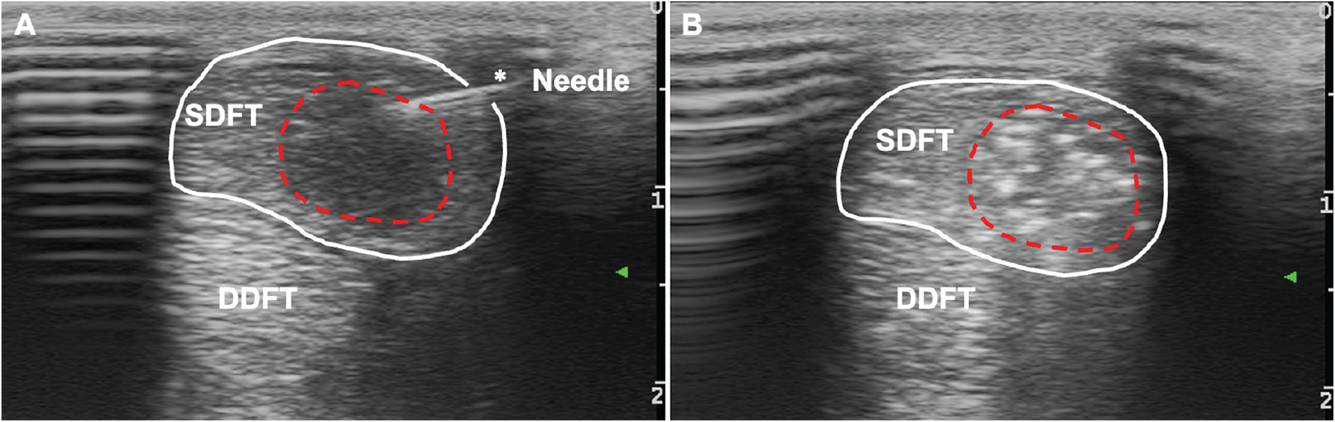
The purpose of this manuscript, which is part of the Currents in One Health series, is to take a comparative approach to stem cell treatment for tendon injury and consider how the horse might inform treatment in other veterinary species and humans. There is increasing experimental and clinical evidence for the use of bone marrow-derived mesenchymal stem cells to treat tendon injuries in the horse. The same evidence does not currently exist for other species. This manuscript will review why the equine superficial digital flexor tendon core lesion might be considered optimal for stem cell delivery and stem cell interaction with the injury environment and will also introduce the concept of stem cell licensing for future evaluation. The companion Currents in One Health by Koch and Schnabel, AJVR, October 2023, addresses in detail what is known about stem cell licensing for the treatment of other diseases using rodent models and how this information can potentially be applied to tendon healing.
Keywords: Achilles tendon; core lesion; mesenchymal stem cell; one health; superficial digital flexor tendon.
Figures
Similar articles
-
How Does Tendon Region, Donor, and the Presence of Disease Affect Protein Composition of the Achilles Tendon?Clin Orthop Relat Res. 2025 Jun 13. doi: 10.1097/CORR.0000000000003578. Online ahead of print. Clin Orthop Relat Res. 2025. PMID: 40522651
-
How to Implement Digital Clinical Consultations in UK Maternity Care: the ARM@DA Realist Review.Health Soc Care Deliv Res. 2025 May;13(22):1-77. doi: 10.3310/WQFV7425. Health Soc Care Deliv Res. 2025. PMID: 40417997 Review.
-
Exploring the application of stem cells in tendon repair and regeneration.Arthroscopy. 2012 Jul;28(7):1018-29. doi: 10.1016/j.arthro.2011.12.009. Epub 2012 Feb 28. Arthroscopy. 2012. PMID: 22381688
-
Adapting Safety Plans for Autistic Adults with Involvement from the Autism Community.Autism Adulthood. 2025 May 28;7(3):293-302. doi: 10.1089/aut.2023.0124. eCollection 2025 Jun. Autism Adulthood. 2025. PMID: 40539213
-
Equine non-septic tenosynovitis: A systematic literature review of site-specific pathological lesions, outcomes and surgical complications.Equine Vet J. 2024 Sep;56(5):842-857. doi: 10.1111/evj.14000. Epub 2023 Sep 21. Equine Vet J. 2024. PMID: 37735927
Cited by
-
The roles and mechanisms of the NF-κB signaling pathway in tendon disorders.Front Vet Sci. 2024 Jun 24;11:1382239. doi: 10.3389/fvets.2024.1382239. eCollection 2024. Front Vet Sci. 2024. PMID: 38978635 Free PMC article. Review.
-
Linkage of jockey falls and injuries with racehorse injuries and fatalities in Thoroughbred flat racing in Victoria, Australia.Front Vet Sci. 2025 Feb 13;11:1481016. doi: 10.3389/fvets.2024.1481016. eCollection 2024. Front Vet Sci. 2025. PMID: 40018508 Free PMC article.
-
Mesenchymal Stem Cells in Veterinary Medicine-Still Untapped Potential.Animals (Basel). 2025 Apr 19;15(8):1175. doi: 10.3390/ani15081175. Animals (Basel). 2025. PMID: 40282009 Free PMC article. Review.
References
-
- Duffy DJ, Beamon WL, Chang YJ, Moore GE. Loop modification of the traditional three-loop pulley pattern improves the biomechanical properties and resistance to 3-mm gap formation in a canine common calcanean teno-osseous avulsion model. Am J Vet Res. 2022;83(8):ajvr.21.09.0139. doi:10.2460/ajvr.21.09.0139 - DOI - PubMed
Publication types
MeSH terms
Grants and funding
LinkOut - more resources
Full Text Sources
