. 2023 Aug 29;6(11):e202301912.
doi: 10.26508/lsa.202301912. Print 2023 Nov.

Grp78 is required for intestinal Kras-dependent glycolysis proliferation and adenomagenesis

Affiliations

Grp78 is required for intestinal Kras-dependent glycolysis proliferation and adenomagenesis

Claudia N Spaan et al. Life Sci Alliance. .

Abstract

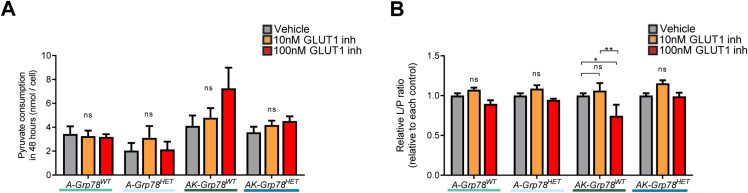
In development of colorectal cancer, mutations in APC are often followed by mutations in oncogene KRAS The latter changes cellular metabolism and is associated with the Warburg phenomenon. Glucose-regulated protein 78 (Grp78) is an important regulator of the protein-folding machinery, involved in processing and localization of transmembrane proteins. We hypothesize that targeting Grp78 in Apc and Kras (AK)-mutant intestines interferes with the metabolic phenotype imposed by Kras mutations. In mice with intestinal epithelial mutations in Apc, Kras G12D and heterozygosity for Grp78 (AK-Grp78 HET ) adenoma number and size is decreased compared with AK-Grp78 WT mice. Organoids from AK-Grp78 WT mice exhibited a glycolysis metabolism which was completely rescued by Grp78 heterozygosity. Expression and correct localization of glucose transporter GLUT1 was diminished in AK-Grp78 HET cells. GLUT1 inhibition restrained the increased growth observed in AK-mutant organoids, whereas AK-Grp78 HET organoids were unaffected. We identify Grp78 as a critical factor in Kras-mutated adenomagenesis. This can be attributed to a critical role for Grp78 in GLUT1 expression and localization, targeting glycolysis and the Warburg effect.


Conflict of interest statement

The authors declare that they have no conflict of interest.

Figures

Figure 1.
Figure 1.. Grp78 heterozygosity reduces colon adenoma initiation and progression in Apc-KrasG12D mice.
(A) Representative images showing the expression of GRP78, KRASG12D, β-catenin by immunohistochemistry and representative images from mRNA in situ hybridization of the floxed Grp78Δ5–7 transcript n consecutive sections. (B) Immunoblot for GRP78 on organoids from indicated genotypes. (C) Representative BrdU staining of indicated genotypes (AK-Grp78WT, AK-Grp78HET). (D) Quantification of BrdU staining in crypts of colon in indicated genotypes. (AK-Grp78WT N = 5, AK-Grp78HET N = 5). (E) Kaplan–Meier curve of prolapse-free survival (A-Grp78WT N = 3, AK-Grp78WT N = 5, AK-Grp78HET N = 5). (F) Representative images of rectal prolapse. (G) Representative whole-mount images of colons. (H) Representative H&E images of entire colons, adenomas are indicated by boxes. (I) Number of colon adenomas (A-Grp78WT N = 3, AK-Grp78WT N = 5, AK-Grp78HET N = 5). (J) Number of colon adenomas separated by size. Small adenoma = <3 crypts, large adenoma = ≥3 crypts (A-Grp78WT N = 3, AK-Grp78WT N = 5, AK-Grp78HET N = 5). *P < 0.05, **P < 0.01, ***P < 0.001; ns, non-significant; ND, Not detectable.
Figure S1.
Figure S1.. Macroscopic and microscopic analyses of intestines of the mice experiment.
(A) Representative caspase staining of indicated genotypes (AK-Grp78WT, AK-Grp78HET) with a quantification of mean caspase+ve cells per 100 crypts. (B) Weight curves of the indicated genotypes (A-Grp78WT N = 3, AK-Grp78WT N = 5, AK-Grp78HET N = 5). (C) Number of small intestinal adenomas (AK-Grp78WT N = 5, AK-Grp78HET N = 5). (D) Number of small intestinal adenomas separated based on size. Small adenoma ≤2 mm, large adenoma >2 mm (AK-Grp78WT N = 5, AK-Grp78HET N = 5). *P < 0.05, **P < 0.01, ***P < 0.001; ns, nonsignificant.
Figure 2.
Figure 2.. Grp78 is critical for Kras-dependent growth in the context of Apc mutant organoids.
(A) Representative bright-field images of Apcfl/flKrasG12D/+Grp78+/+ and Apcfl/flKrasG12D/+Grp78+/fl small intestinal organoids upon recombination with 4-OHT (AK-Grp78WT and AK-Grp78HET) or treatment with the vehicle. (B) Organoid growth measured by the surface area of Apcfl/flKrasG12D/+Grp78+/+ and Apcfl/flKrasG12D/+Grp78+/fl organoids upon recombination with 4OHT (AK-Grp78WT and AK-Grp78HET) or treatment with the vehicle. N = 3 per genotype. (C) Representative bright-field images of A-Grp78WT, A-Grp78HET, AK-Grp78WT, and AK-Grp78HET organoids. Mice with genotype VillinCreERT2, Apcfl/fl, KrasG12D/+ or +/+ and Grp78+/+ or +/fl were crossed to gain four different types of organoids. (D) Growth of organoids 72 h after induction of the genotypes. Quantification of area relative to the control A-Grp78WT. (E) Proliferation measured by EdU incorporation relative to the control A-Grp78WT. (F) Quantitative RT–PCR analysis with specific primers for the Grp78Δ5–7 mRNA. (G) Global protein translation measured with [35S]methionine incorporation in organoids from the indicated genotypes. (H) Quantitative RT–PCR analysis for Lgr5 and Olfm4 in organoids from the indicated genotypes. (I) Bright-field image of organoids grown after single-cell seeding. (J) Quantification of organoids grown from equal numbers of single cells from indicated genotypes. Quantification of area relative to the control AK-Grp78WT. *P < 0.05, **P < 0.01, ***P < 0.001; ns, nonsignificant.
Figure S2.
Figure S2.. Intestinal organoids show increased ER stress upon Grp78 heterozygosity.
(A, B) Quantitative RT–PCR analysis for Grp78 (A) and Grp94 (B) mRNA on AK-Grp78WT and AK-Grp78HET organoids. *P < 0.05, **P < 0.01, ***P < 0.001; ns, nonsignificant.
Figure 3.
Figure 3.. GLUT1-dependent glycolysis in Apc-Kras mutant organoids is regulated by Grp78.
(A) Oxygen consumption rate in organoids of indicated genotypes, measured by Agilent Seahorse. (B) Basal respiration, ATP-linked respiration, and maximum respiration in the indicated organoids. (C) Glycolytic function in the organoids, measured by Agilent Seahorse. (D) Glycolysis and glycolytic capacity in the indicated organoids. (E) Grp78 mRNA and Glut1 mRNA expressions derived from microarray analysis of indicated mouse organoids (GSE143509). (F) Immunoblots of GRP78 and GLUT1 from indicated organoids. (G) Flow cytometric immunostaining of membrane-localised GLUT1 protein on live cells, percentage Alexa 647+ve of the parent (single cells, PI negative). (H) Glucose consumption upon treatment with vehicle, 10 , and 100 nM of GLUT1-inhibitor BAY-876. (I) Lactate production upon treatment with vehicle, 10 , and 100 nM of GLUT1-inhibitor BAY-876. (J) EdU incorporation upon treatment with vehicle, 10 , and 100 nM of GLUT1-inhibitor BAY-876. (K) Quantitative RT–PCR analysis for Pgc1a mRNA on organoids of indicated genotypes upon treatment with vehicle, 10 , and 100 nM of GLUT1-inhibitor BAY-876. *P < 0.05, **P < 0.01, ***P < 0.001, ns, nonsignificant.
Figure S3.
Figure S3.. GLUT1 is up-regulated in human intestinal adenomas.
(A) GRP78 mRNA and GLUT mRNA expression derived from microarray analysis of indicated human colon normal colonic mucosa and colon tumors (mutation status unknown) (GSE33114). (B) Immunoblot of the protein level of GLUT1 in human colonic tissue, normal epithelium versus carcinoma tissue. 10 pairs are showed. Quantification of protein expression was calculated using ImageJ, relative expression of carcinoma tissue compared with normal adjacent tissue.
Figure S4.
Figure S4.. Gating strategy and raw data of GLUT1 immunostaining on organoids.
(A) Gating strategy of single-cell organoids after incubating with GLUT1 antibody, second anti-rabbit Alexa Fluor 647, and PI for determining live cells. (B) Raw data of control samples, which lacked first and/or second antibodies. (C) Representative gating of GLUT1 immunostaining on the described organoids. PI, Propidium iodide.
Figure S5.
Figure S5.. Effects of GLUT1 inhibition, Grp78, and Kras on pyruvate/lactate metabolism.
(A) Pyruvate consumption in 48 h after adding the vehicle, 10 nM GLUT1 inhibitor or 100 nM GLUT1 inhibitor in the A-Grp78WT, A-Grp78HET, AK-Grp78WT, and AK-Grp78HET organoids. (B) Lactate/Pyruvate ratio in the above described organoids. *P < 0.05, **P < 0.01, ***P < 0.001; ns, nonsignificant.

References

    1. Bennecke M, Kriegl L, Bajbouj M, Retzlaff K, Robine S, Jung A, Arkan MC, Kirchner T, Greten FR (2010) Ink4a/Arf and oncogene-induced senescence prevent tumor progression during alternative colorectal tumorigenesis. Cancer cell 18: 135–146. 10.1016/j.ccr.2010.06.013 - DOI - PubMed
    1. Bertolotti A, Zhang Y, Hendershot LM, Harding HP, Ron D (2000) Dynamic interaction of BiP and ER stress transducers in the unfolded-protein response. Nat Cell Biol 2: 326–332. 10.1038/35014014 - DOI - PubMed
    1. Bos JL, Fearon ER, Hamilton SR, Verlaan-de Vries M, van Boom JH, van der Eb AJ, Vogelstein B (1987) Prevalence of RAS gene mutations in human colorectal cancers. Nature 327: 293–297. 10.1038/327293a0 - DOI - PubMed
    1. Chang JC, Go S, Gilglioni EH, Duijst S, Panneman DM, Rodenburg RJ, Li HL, Huang HL, Levin LR, Buck J, et al. (2021) Soluble adenylyl cyclase regulates the cytosolic NADH/NAD(+) redox state and the bioenergetic switch between glycolysis and oxidative phosphorylation. Biochim Biophys Acta Bioenerg 1862: 148367. 10.1016/j.bbabio.2020.148367 - DOI - PubMed
    1. Colnot S, Niwa-Kawakita M, Hamard G, Godard C, Le Plenier S, Houbron C, Romagnolo B, Berrebi D, Giovannini M, Perret C (2004) Colorectal cancers in a new mouse model of familial adenomatous polyposis: Influence of genetic and environmental modifiers. Lab Invest 84: 1619–1630. 10.1038/labinvest.3700180 - DOI - PubMed

Publication types

LinkOut - more resources