. 2023 Aug 29;14(1):5253.
doi: 10.1038/s41467-023-40760-6.

Loss of LCMT1 and biased protein phosphatase 2A heterotrimerization drive prostate cancer progression and therapy resistance

Affiliations

Loss of LCMT1 and biased protein phosphatase 2A heterotrimerization drive prostate cancer progression and therapy resistance

Reyaz Ur Rasool et al. Nat Commun. .

Abstract

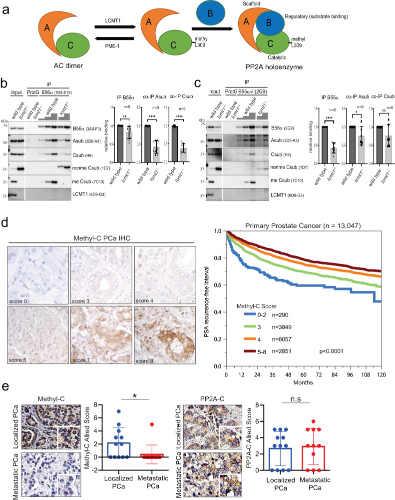
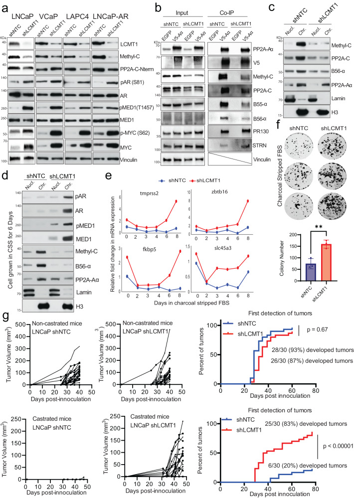
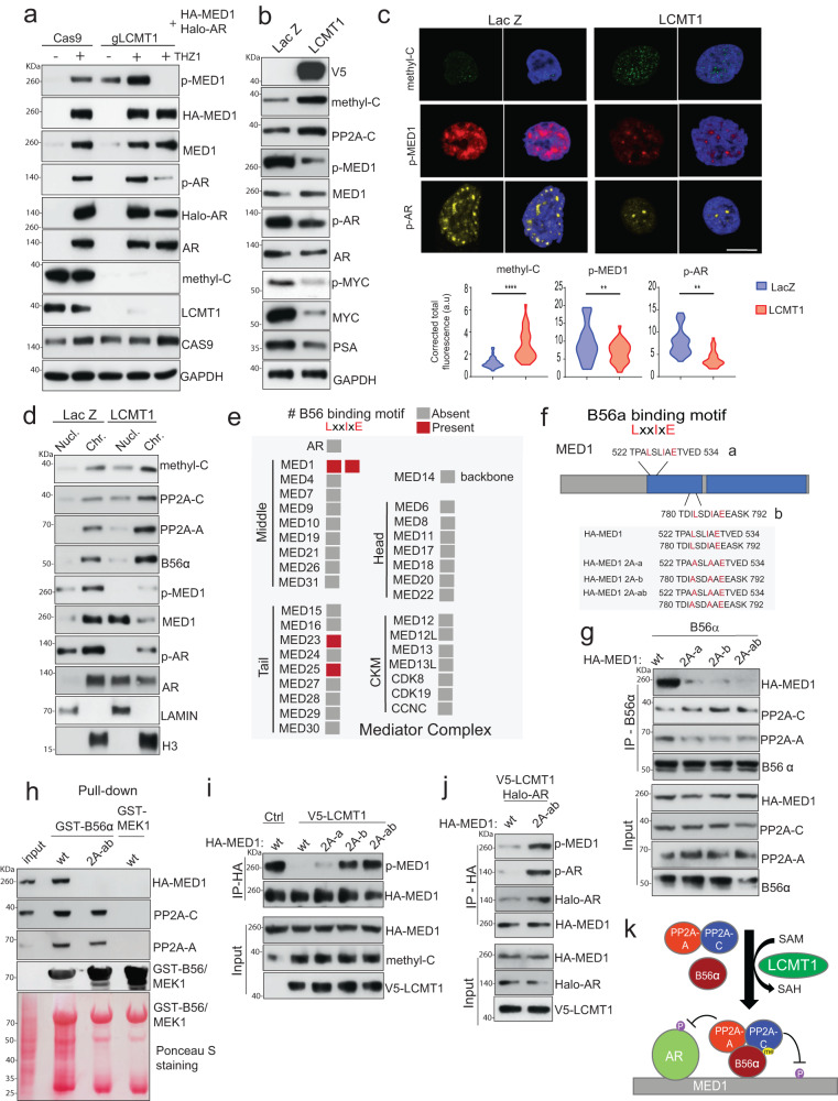
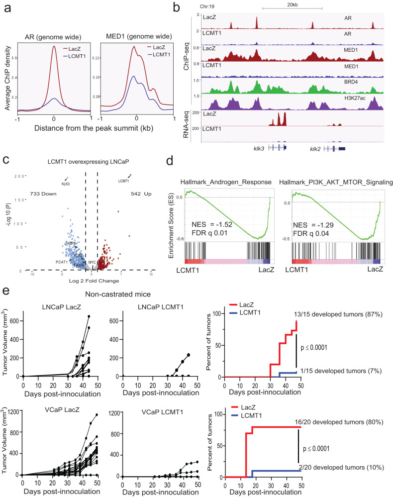
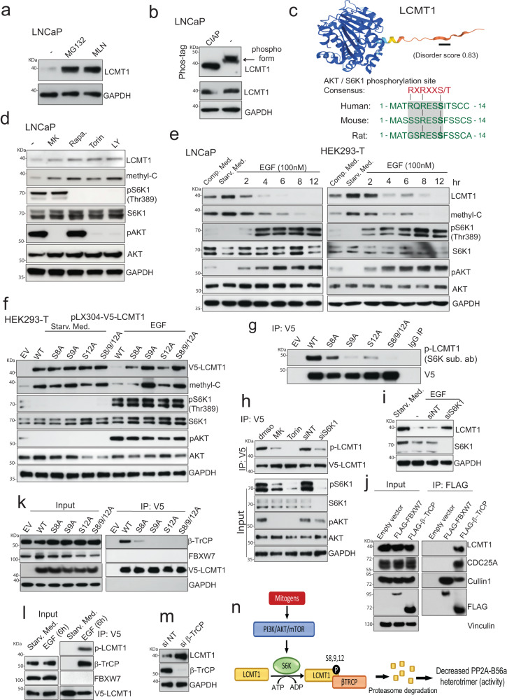
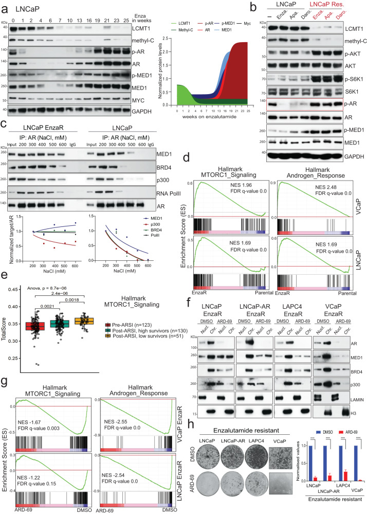
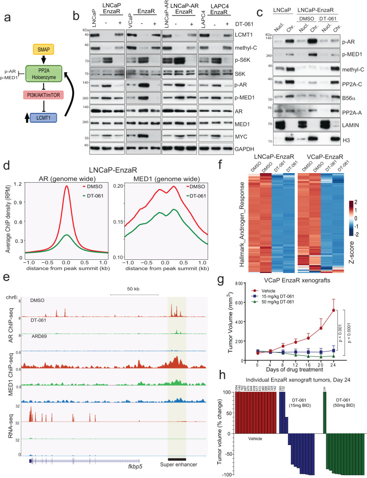
Loss of the tumor suppressive activity of the protein phosphatase 2A (PP2A) is associated with cancer, but the underlying molecular mechanisms are unclear. PP2A holoenzyme comprises a heterodimeric core, a scaffolding A subunit and a catalytic C subunit, and one of over 20 distinct substrate-directing regulatory B subunits. Methylation of the C subunit regulates PP2A heterotrimerization, affecting B subunit binding and substrate specificity. Here, we report that the leucine carboxy methyltransferase (LCMT1), which methylates the L309 residue of the C subunit, acts as a suppressor of androgen receptor (AR) addicted prostate cancer (PCa). Decreased methyl-PP2A-C levels in prostate tumors is associated with biochemical recurrence and metastasis. Silencing LCMT1 increases AR activity and promotes castration-resistant prostate cancer growth. LCMT1-dependent methyl-sensitive AB56αCme heterotrimers target AR and its critical coactivator MED1 for dephosphorylation, resulting in the eviction of the AR-MED1 complex from chromatin and loss of target gene expression. Mechanistically, LCMT1 is regulated by S6K1-mediated phosphorylation-induced degradation requiring the β-TRCP, leading to acquired resistance to anti-androgens. Finally, feedforward stabilization of LCMT1 by small molecule activator of phosphatase (SMAP) results in attenuation of AR-signaling and tumor growth inhibition in anti-androgen refractory PCa. These findings highlight methyl-PP2A-C as a prognostic marker and that the loss of LCMT1 is a major determinant in AR-addicted PCa, suggesting therapeutic potential for AR degraders or PP2A modulators in prostate cancer treatment.


Conflict of interest statement

G.N. has an equity interest in RAPPTA Therapeutics and serves as consultants, along with C.O. E.O. serves as a consultant to Millipore Corporation and RAPPTA Therapeutics and has option interests in RAPPTA Therapeutics. The Medical University of Vienna and the Regents of The University of Michigan on behalf of the authors EO and GN have filed patent WO2021148681A1 on the PP2A methyl-C subunit specific monoclonal antibody 7C10-C5 with the title: Antibodies specifically binding the carboxymethylated catalytic subunit of protein phosphatase 2A. All other authors declare no competing interests.

Figures

Fig. 1
Fig. 1. Methyl-PP2A-C loss is associated with prostate cancer progression.
a Schematic showing methylation-sensitive PP2A holoenzyme complex. b, c Reduced levels of B56 and B55 subunit-containing holoenzymes in cells lacking PP2A methylation as a result of CRISPR mediated knockout of LCMT1. Immunoblotting of lysates and anti-B56α or anti-B55α/δ immunoprecipitates from lysates of HAP1 wildtype and lcmt1−/− cells. 1/10 of the input lysate was loaded for the B56α blot, and 1/160 of the input lysate for all other blots. The panels originate from 3 independent blotting membranes, which were sequentially incubated with the indicated antibodies. The blots are representative of n = 9 (B56α) and n = 6 (B55α/δ) independent immunoprecipitation experiments. The amounts of immunoprecipitated B56α and B55α/δ were quantified, and the coimmunoprecipitated A and C subunit levels were normalized to the B56α or B55α/δ levels, which were set to 1 for the wild-type cells. Statistical significance of quantified protein levels was assessed using an unpaired two-sided Student’s t-test. b **P = 0.0048 ****P < 0.0001 c ****P < 0.0001 *P = 0.0434, ns P = 0.0932. Data are presented as mean values ± s.d. d Representative images of methyl-C immunohistochemistry in primary prostate cancers. From upper left to lower right: score 0, 3, 4, 5, 7, and 8. A low immunohistochemistry methyl-C score is linked to early biochemical recurrence in the tissue microarray cohort of 13,047 prostate cancers with available follow-up data. e Representative immunohistochemistry images showing expression of methyl-PP2A-C (methyl-C) and total PP2A-C in localized and metastatic prostate cancer tissues. Bar graphs show the quantification, error bars mean ± s.d., n = 11 metastatic tissue and n = 12 localized tissue samples. Student two-tailed t-test, p value: 0.0376 (methyl-C), p value: 0.7902 (PP2A-C). *P < 0.05, **P < 0.01, ***P < 0.001 ***P < 0.0001, ns non-significant.
Fig. 2
Fig. 2. LCMT1 loss activates AR signaling and promotes castration-independent tumor growth.
a Methyl-C reduction upon LCMT1 silencing results in a concomitant increase in p-AR, p-MED1, and MYC expression. Representative immunoblots in a panel of prostate cancer cells stably expressing shNTC or shLCMT1 showing indicated proteins. Vinculin was used as a loading control. b Reduced AB56αCme and AB55αCme (PP2A) heterotrimers in LCMT1 silenced LNCaP cells. Coimmunoprecipitation (IP) analysis using lysates from the indicated cells with V5-PP2A-Aα and EGFP-specific antibodies, demonstrating the interaction between PP2A-Aα, methyl-PP2A-C, B56-α, B55-α. PR130 and STRN are used as methylation insensitive controls and EGFP as a negative IP control. Total lysate was used as input control. c LCMT1 loss disrupts canonical AB56αCme heterotrimer association with chromatin. Chromatin and soluble nuclear fractions from control and LCMT1 silenced LNCaP cells were subjected to immunoblotting for the indicated proteins. Lamin and H3 served as controls for the nuclear and chromatin fractions, respectively. d LCMT1 abrogation resists androgen-ablation arbitrated suppression of AR and MED1 recruitment to the chromatin. Chromatin and soluble nuclear fractions extracted from the cells grown in charcoal-stripped fetal bovine serum (CSS) medium for six days were subject to immunoblotting for the indicated proteins. Lamin and H3 served as controls for nuclear and chromatin fractions, respectively. e Continuous AR transcriptional activity under androgen deprivation. qRT-PCR for AR-regulated genes in cells grown in CSS-containing medium. f Colony formation assay demonstrates the increased proliferation of LCMT1 depleted cells in hormone-deprived media. Cells were cultured in CSS media for 10 days, followed by crystal violet staining (top), and quantification (bottom), error bars mean ± s.d., n = 3 biologically independent replicates. Students two-tailed t-test, p value: 0.006. g Castration-independent tumor growth in vivo upon LCMT1 silencing. Castrated and non-castrated mice bearing shLCMT1- or shNTC-LNCaP were evaluated for tumor growth. The tumor volume is plotted (left) and the percentage of tumors first detected is shown (right). Fisher’s exact test of relative risk. The data in ad are representative of n = 3 independent experiments.
Fig. 3
Fig. 3. LCMT1 abrogates AR-MED1 transcriptional activity by stabilizing the AB56αCme heterotrimer on chromatin.
a LCMT1 loss leads to the accumulation of hyperphosphorylated AR and MED1. Cas9 control and LCMT1 null HEK293-T cells were co-transfected with HA-MED1 and Halo-AR for 48 h or treated with 100 nM THZ1 (CDK7i) for 12 h followed by immunoblotting for the indicated proteins. b LCMT1 overexpression increases methyl-C levels with a concomitant reduction in AR-MED1 phosphorylation. Protein lysates were subjected to immunoblotting for the indicated proteins. c Representative confocal images show nuclear methyl-C expression is inversely correlated with p-MED1 and p-AR expression (top). Quantification (bottom) is in the form of corrected total cell fluorescence, with two-tailed t-tests *p < 0.05, **p < 0.01. Scale bar: 10 μm. d Immunoblotting showing enhanced recruitment of AB56αCme and loss of AR and MED1 on the chromatin upon LCMT1 overexpressing. Lamin and H3 served as controls. e Color-coded plot showing the presence or absence of B56α binding SLiMs on AR and subunits of human Mediator complex. f Schematic depicting two B56α binding motifs on MED1. The blue region indicates IDR – intrinsically disordered region. MED1 mutants used in the study are shown. g MED1 binds PP2A-B56α through two conserved LxxIxE motifs. B56α (B56a) was co-transfected with either HA-MED1-wild type (wt) or the indicated 2A mutant constructs in HEK293-T cells for 72 h post transfection, cell lysates were subjected to co-IP with the B56α antibody for the shown proteins. h GST-pull-down assay showing purified GST-B56α binds to MED1-wt not 2A-ab mutant. GST-MEK1 was used as a negative control. i HEK293-T cells were transfected with either MED1-wt or the indicated MED1 mutant alone or with pLX304-V5-LCMT1 for 72 h, followed by co-IP and immunoblotting for the indicated proteins. j Dephosphorylation of AR by PP2A-B56α is through MED1 binding. Co-IP with HA antibody was performed using lysates prepared from HEK293-T cells transfected with Halo-AR and V5-LCMT1 constructs and either HA-MED1-wt or HA-MED1-2A-ab. k Schematic showing LCMT1 mediated PP2A-B56α heterotrimerization directly targets MED1-AR phosphorylation. The data in a, b, d, gj are representative of at least n = 3 independent biological replicates.
Fig. 4
Fig. 4. LCMT1 abrogates AR-MED1 transcriptional activity and prostate cancer growth.
a LCMT1 over-expression decreases AR and MED1 recruitment to the chromatin. Genome-wide averaged AR and MED1 ChIP-seq enrichment profile plot in LNCaP cells stably overexpressing LCMT1 or LacZ. b Genome browser tracks of AR and MED1 binding at klk2 and klk3 loci in the indicated cells, and BRD4/H3K27ac tracks from LNCaP cells. The tracks at the bottom show the RNA-seq gene expression. c Volcano plot of RNA-seq results showing differentially expressed genes in LCMT1 versus LacZ expressing LNCaP cells. d GSEA plots showing negative enrichment of Hallmark Androgen Response and PI3K/AKT/mTOR pathway genes in LCMT1 overexpressing LNCaP cells. NES normalized enrichment score, FDR false discovery rate. e LCMT1 suppresses in vivo tumor growth. Naive mice bearing either LNCaP or VCaP-LCMT1 and -LacZ xenografts were evaluated for tumor growth for the given number of days. The tumor volume (measured twice per week using calipers) and the percentage of tumors detected is shown. P value indicates Fisher’s exact test of relative risk.
Fig. 5
Fig. 5. Phosphorylation-dependent degradation of LCMT1 mediated by S6K1- and β-TrCP.
a Immunoblotting showing MG132 and MLN4924 (10 µM, 12 h) restore LCMT1 protein expression. b Phos-tag analysis showing LCMT1 phosphorylation. Control immunoblots were done on standard gels. c The LCMT1 AlphaFold-predicted structure with AKT/S6K1 phosphorylation site is indicated with a black line, and the disorder score is shown. Position and cross-species sequence conservation is shown below. d Cells were treated with vehicle, MK: MK-2206 (AKTi) 1 µM, Rapa: Rapamycin (mTORi) 10 µM, Torin1 (mTORi) 0.1 µM or LY: LY294002 (PI3Ki) 15 µM, for 24 h followed by immunoblotting. e Rapid degradation of LCMT1 in response to mitogens. Cells were either grown in complete medium or serum-starved for 24 h and then stimulated with EGF followed by immunoblotting. f Twenty-four hours post transfection with the indicated constructs, the cells were serum starved for 24 h and then either stimulated with EGF or continued to grow in the absence of serum for another 24 h followed by immunoblotting. g Forty-eight hours post transfection with the indicated constructs, protein extract was used for V5 pull-down and probed with p-AKT/S6K-substrate (p-LCMT1) or V5 antibodies. h S6K1 phosphorylates LCMT1. HEK293-T were transfected with V5-LCMT1 and treated with indicated drugs or siNT/siS6K1. Protein extract was used for the V5 pull-down and probed for indicated proteins. i S6K1 silencing restores LCMT1. HEK293-T cells were serum-starved and transfected with siS6K1 followed by EGF stimulation and then immunoblotting. j LCMT1 and β-TrCP interact directly. HEK293-T were transfected with constructs encoding an empty vector or FLAG-tagged FBXW7 or FLAG-tagged β-TrCP. Forty-eight hours post transfection, the cells were treated with 5 μM MLN4924 for 6 h followed by co-IP and immunoblotting for the indicated proteins. k HEK293-T cells were transfected with the indicated constructs. Transfected cells were then grown in the presence of 5 μM MLN4924 for 6 h followed by co-IP with anti-V5 and immunoblotting with indicated antibodies. l LCMT1 phosphorylation is required for its interaction with β-TrCP. HEK293-T cells were transfected with the V5-LCMT1 construct. Twenty-four hours post transfection, the cells were serum-starved for 24 h, followed by EGF stimulation for 6 h. Protein extract was used for co-IP with anti-V5 and probed for indicated proteins. m HEK293-T cells were transfected with si-β-TrCP followed by immunoblotting. n Schematic depicting S6K-induced phosphorylation and β-TrCP mediated degradation of LCMT1. All the experiments were repeated at least three times independently with similar observation.
Fig. 6
Fig. 6. AR addiction is associated with reduced LCMT1 and hyperactive AKT/S6K1 signaling in enzalutamide refractory (EnzaR) PCa cells.
a Evolution of EnzaR state is associated with gradual loss of LCMT1 and Leucine-309 methylation. LNCaP cells grown in the presence of enzalutamide were harvested at the indicated weeks, and the lysates prepared were immunoprobed for the indicated proteins. Normalized densitometric (ImageJ) plot for proteins is shown. The experiment was performed in two separate cell lines with the similar observations. b EnzaR, ApaR, and DaroR cells show reduced LCMT1 expression with a concomitant increase in p-AKT, p-S6K1, p-AR, and p-MED1. Representative immunoblots from n = 3 independent experiments show the effect on the indicated proteins by the transient (24 h) and chronic exposure with 10 µM of the indicated anti-androgens. c Top: AR co-IP was performed with increasing salt concentrations in washing buffer followed by the immunoblotting for the indicated proteins. Bar graphs show the normalized ratio of the target protein to AR. The data is representative of n = 2 biological replicates. d GSEA plot showing enrichment of the indicated msigDB signatures in EnzaR cells. e Box plot showing gene signature scores for Hallmark MTORC1 signaling in pre-ARSI therapy and post-ARSI therapy stratified by overall survival from biopsy. p values calculated from One-way Anova are shown. The center line shows the median, the box limits show the 75th and 25th percentiles and the whiskers show minimum-maximum values. f Loss of chromatin-bound AR, MED1, and BRD4 upon ARD-69 (AR-PROTAC) treatment. Chromatin and soluble nuclear fractions from cells treated with 100 nM ARD-69 for 12 h were probed for the indicated proteins. The data is representative of n = 3 biological replicates. g GSEA plot showing negative enrichment of the indicated signatures in EnzaR cells treated with ARD-69. h Indicated cell lines were cultured either in the presence of vehicle or 100 nM ARD-69 for 12–14 days, followed by crystal violet staining (n = 3). In the case of VCaP -representative bright field images are shown. Normalized quantification of crystal violet stain/cell viability is shown. Error bar represents the mean ± s.d. (n = 3). Statistical significance as calculated by two-tailed t-test is represented as ****p < 0.0001.
Fig. 7
Fig. 7. SMAP induced feedforward activation of LCMT1 attenuates AR addiction in enzalutamide refractory cells.
a Schematic showing the feedforward effect of DT-061 on PP2A holoenzyme activation and LCMT1 stabilization. b DT-061 restores LCMT1 protein expression by reducing pS6K, resulting in lower p-AR and p-MED1 levels. Protein lysates were prepared from the EnzaR cells treated with vehicle or 10μM DT-061 for 24 h, followed by protein extraction and immunoblotting for the indicated protein. Lysates from the respective parental lines served as controls. GAPDH was used as the loading control. c DT-061 stabilizes the PP2A-B56α complex on the chromatin resulting in the loss of chromatin-bound p-AR and p-MED1. Chromatin and nuclear fractions from parental LNCaP and LNCaP-EnzaR cells treated with either DMSO or 10μM DT-061 for 12 h were used to probe the indicated proteins. Lamin and total H3 served as controls for the soluble nuclear and chromatin fractions, respectively. d DT-061 decreases genome-wide AR and MED1 binding. Genome-wide averaged AR and MED1 ChIP-seq enrichment in LNCaP-EnzaR cells upon treatment with 10μM DT-061 for 12 h. e Genome browser tracks of AR and MED1 binding at fkbp5 locus in the indicated condition for LNCaP-EnzaR cells. The superenhancer associated with this region is indicated with a black bar. The tracks at the bottom show the fkbp5 transcript expression at this corresponding locus. f Heatmaps of FPKM z-score displaying negative enrichment of msigDB Hallmark Androgen response signature genes in LNCaP EnzaR and VCaP-EnzaR treated with vehicle or 10 μM DT-061 for 24 h. g DT-061 blocks enzalutamide refractory prostate cancer growth in vivo. Mice bearing VCaP-EnzaR xenografts received vehicle (n = 12) or 15 mg/kg (n = 12) or 50 mg/kg (n = 15) DT-061 twice daily (b.i.d) for 3 weeks. Tumor volume was measured twice per week using calipers. Data are mean ± s.e.m., and *p values indicated were computed using unpaired two-tailed t-test. h Percent change in volume for each tumor after 24 days of treatment is shown as a waterfall plot (y-axis). For b and c the experiments were repeated at least two times independently with similar observations.
Fig. 8
Fig. 8. Model showing the S6K-LCMT1-PP2A-AR axis in normal prostate and advanced refractory prostate cancer.
Methyl-sensitive PP2A holoenzyme complex is destabilized by PI3K/AKT/mTOR pathway-mediated degradation of LCMT1 through β-TRCP, resulting in hyperphosphorylated chromatin-bound stable AR transcriptional complex. Biased heterotrimerization of PP2A by small molecular activators of phosphatase lead to a feedforward LCMT1 stabilization via AKT-S6K1 inhibition, resulting in dephosphorylation of the AR transcriptional complex and death of refractory AR-addicted cells. The schematic was created with BioRender.com.

References

    1. Hanahan D, Weinberg RA. The hallmarks of cancer. Cell. 2000;100:57–70. - PubMed
    1. Brautigan DL, Shenolikar S. Protein serine/threonine phosphatases: keys to unlocking regulators and substrates. Annu. Rev. Biochem. 2018;87:921–964. - PubMed
    1. Shi Y. Serine/threonine phosphatases: mechanism through structure. Cell. 2009;139:468–484. - PubMed
    1. Sangodkar J, et al. All roads lead to PP2A: exploiting the therapeutic potential of this phosphatase. FEBS J. 2016;283:1004–1024. - PMC - PubMed
    1. Eichhorn PJ, Creyghton MP, Bernards R. Protein phosphatase 2A regulatory subunits and cancer. Biochim Biophys. Acta. 2009;1795:1–15. - PubMed

Publication types