. 2023 Aug 29;13(1):14158.
doi: 10.1038/s41598-023-40638-z.

2-Photon imaging of fluorescent proteins in living swine

Affiliations

2-Photon imaging of fluorescent proteins in living swine

Beth A Costine-Bartell et al. Sci Rep. .

Abstract

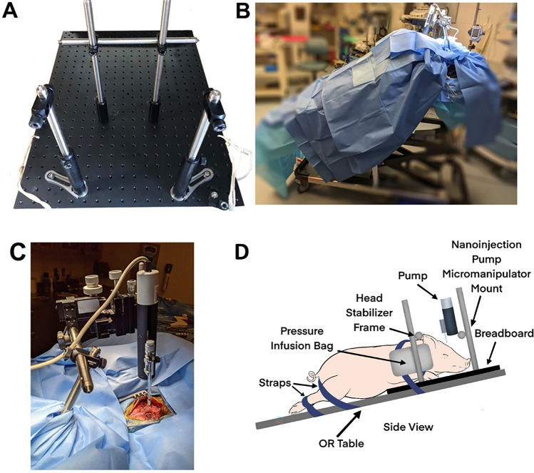
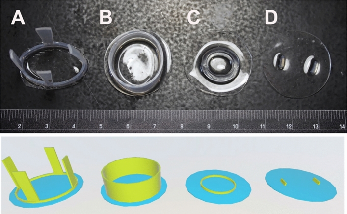
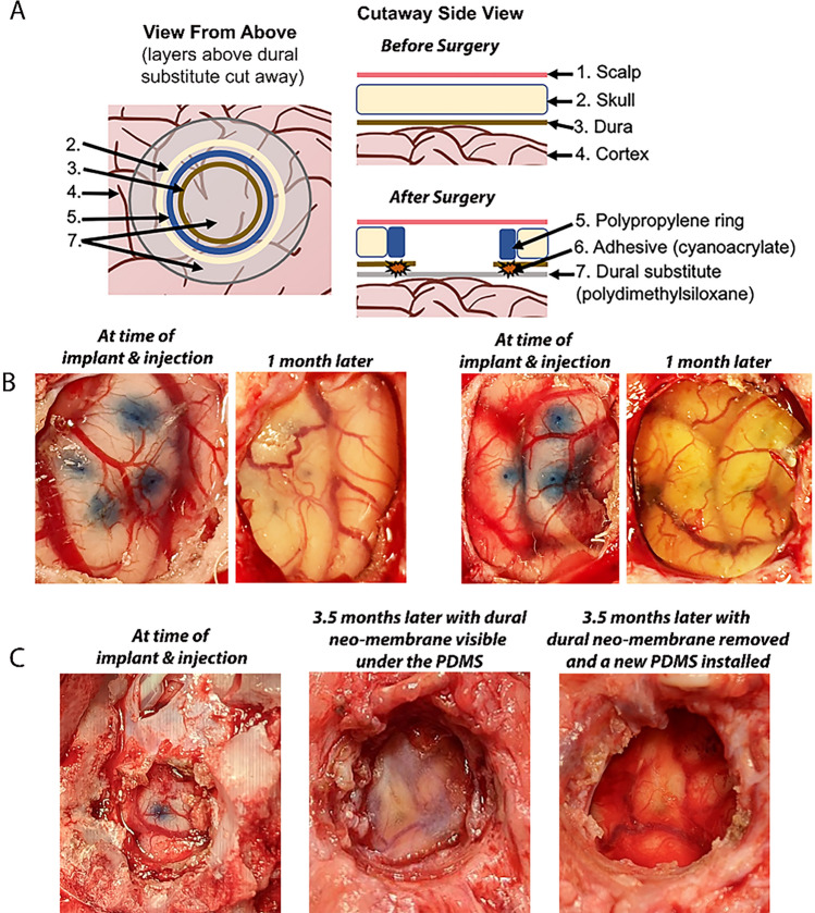
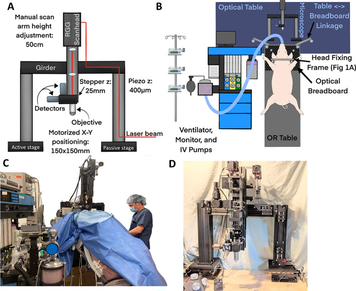
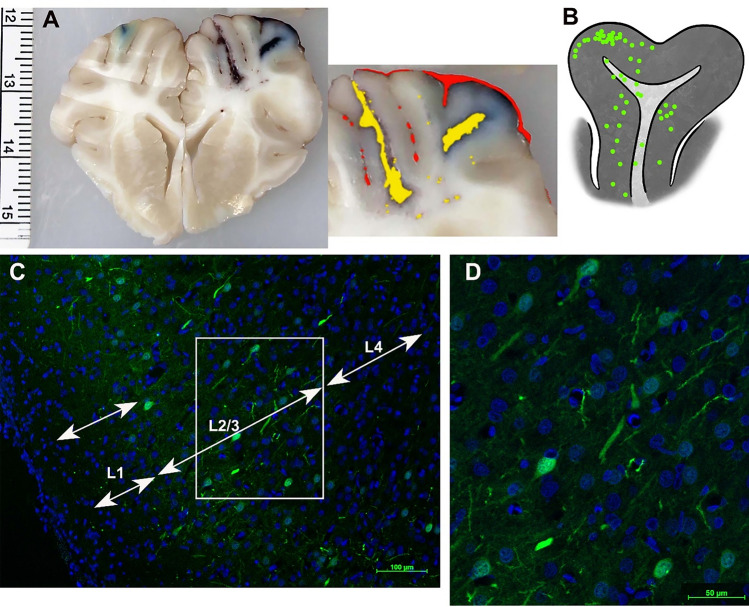
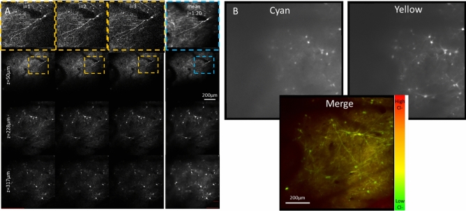
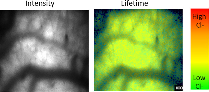
A common point of failure in translation of preclinical neurological research to successful clinical trials comes in the giant leap from rodent models to humans. Non-human primates are phylogenetically close to humans, but cost and ethical considerations prohibit their widespread usage in preclinical trials. Swine have large, gyrencencephalic brains, which are biofidelic to human brains. Their classification as livestock makes them a readily accessible model organism. However, their size has precluded experiments involving intravital imaging with cellular resolution. Here, we present a suite of techniques and tools for in vivo imaging of porcine brains with subcellular resolution. Specifically, we describe surgical techniques for implanting a synthetic, flexible, transparent dural window for chronic optical access to the neocortex. We detail optimized parameters and methods for injecting adeno-associated virus vectors through the cranial imaging window to express fluorescent proteins. We introduce a large-animal 2-photon microscope that was constructed with off-the shelf components, has a gantry design capable of accommodating animals > 80 kg, and is equipped with a high-speed digitizer for digital fluorescence lifetime imaging. Finally, we delineate strategies developed to mitigate the substantial motion artifact that complicates high resolution imaging in large animals, including heartbeat-triggered high-speed image stack acquisition. The effectiveness of this approach is demonstrated in sample images acquired from pigs transduced with the chloride-sensitive fluorescent protein SuperClomeleon.


Conflict of interest statement

The authors declare no competing interests.

Figures

Figure 1
Figure 1
Surgery for cortical AAV injection and installation of the cranial imaging window. (A) The pig’s head was secured in the breadboard with two vertical bars next to their snout and the horizontal bar pressing on the dorsal portion of its snout. Pressure infusion bags were inflated between the pig’s head and the back two vertical bars with the area between the chest and front legs pressed into the vertical bars. The pig’s front legs were tied to the board and the board tied to the OR table. Once the pig was draped, a sterile bar was placed horizontally between the back two vertical bars. (B) Photograph of the pig in position for AAV injection and imaging port implant. The pig was placed at a 30–45° angle to encourage the brain to fall away from the dura for implantation of PDMS avoiding cortical damage. (C) The sterile cross bar added to the vertical bars after draping the animal. Then, the sterile injector and manipulator were mounted to the cross bar. (D) Schematic depiction of pig positioning shown in (B).
Figure 2
Figure 2
The evolution of the dural substitute (and corresponding 3D renderings). (A) PDMS with silicone straps worked well but prevented later cortical impact. (B) PDMS with “top hat” portion to fill the edges of the burr hole. This version was difficult to place without causing cortical trauma. (C) PDMS with flat silicone ring. The ring didn’t provide much advantage and limited the area for 2P imaging. (D) Flat PDMS with ridges to identify the center of the PDMS. This iteration was relatively easy to install, allowed centering of the PDMS, and could withstand injections and cortical impact.
Figure 3
Figure 3
Cranial imaging window. (A) The cranial imaging window system from the top and the side that prevented the adhesion of the healing of the dura onto the cortex that obfuscated the surface of the cortex as well as tissue growth from the burr hole of the skull. (B) Two burr holes displaying the PDMS overlaying the cortex immediately after AAV injection and the pristine window 1 month later. (C) The cranial window at installment, after 3.5 months with dural neomembrane obscuring the cortex, and immediately after dural neo-membrane removal and PDMS re-installation. This iteration of the cranial window had an adjacent subdural electrode strip that entered posterior to the imaging window.
Figure 4
Figure 4
2-photon rig and anesthetic support set up. (A) Schematic depiction of key elements of the microscope body, highlighting the large range of motion for accommodating a large animal. (B) Schematic of room layout during an imaging session. (C) Photograph of an imaging experiment with an 84 kg pig under the 2-photon microscope. The surgery table was resting on and mechanically coupled to the optical table. The ventilator is placed perpendicular to the optical table to the left of the pig with the ECG, gas analyzers, physiology monitor (not shown) and IV pumps on the ventilator (scale: human = 1.7 m). (D) Photograph of overall microscope construction.
Figure 5
Figure 5
Transduction of the cortex with SuperClomeleon. (A) Coronal slab of brain with bilateral injections of SuperClomeleon AAV into the rostral gyrus, 5 weeks after injection and 5 h after unilateral cortical impact (right hemisphere). As expected, the cortical impact site is moderately swollen with subarachnoid (marked red in inset) and intraparenchymal hemorrhages (marked yellow in inset) and the injection site visible (blue dye). On the left cortex, the injection site (blue dye) remains pristine following AAV injection and after 2 h of 2P imaging. (B) Schematic demonstrating the distribution of SuperClomeleon+ neurons in the rostral gyrus down to the sulci. SuperClomeleon+ neurons were dense in Layers 2/3 of the gray matter, were dispersed through all layers to Layer 6, and were in the gyral white matter, perhaps expressed by interstitial neurons in pig. This was pig 86.027 (Supplemental Fig. 1), where conditions were optimized and repeated for several pigs and results consistent. (C) Low power (10×) photomicrograph of SuperClomeleon+ neurons (green) with nuclei (DAPI, blue; scale bar = 100 μM). SuperClomeleon+ neurons are dense in Layer 2/3 and dispersed in Layer 4. (D) Higher power photomicrograph (20×; scale bar = 50 μM) of Layer 2/3 in Panel (C) (white box) demonstrates SuperClomeleon expression in pyramidal neuron cell bodies and neurites.
Figure 6
Figure 6
Imaging in vivo intraneuronal chloride of the cortex. (A) The heartbeat triggered acquisition of a fast z-stack spanning 600 × 600 × 400 µm provided stable images of the fluorescent protein-transduced neurons in vivo. Because these volumes were acquired between heartbeats, individual iterations (first 3 columns) could be averaged together to increase dynamic range and decrease shot noise in the final image stacks (4th column). (B) Merging the cyan and yellow fluorescent protein signals from SuperClomeleon provides a pseudo-colored imaged of intracellular chloride.
Figure 7
Figure 7
Imaging in vivo extracellular chloride of the cortex. After injection of a 10 kD dextran-linked chloride sensitive organic dye, fluorescence lifetime images were acquired using the heartbeat-triggered volume acquisition described above. This produced high resolution intensity (left) images and lifetime (right) images that can be used to quantify extracellular chloride.

Update of

References

    1. Kola I, Landis J. Can the pharmaceutical industry reduce attrition rates? Nat. Rev. Drug Discov. 2004;3:711–715. doi: 10.1038/nrd1470. - DOI - PubMed
    1. Geerts H. Of mice and men: Bridging the translational disconnect in CNS drug discovery. CNS Drugs. 2009;23:915–926. doi: 10.2165/11310890-000000000-00000. - DOI - PubMed
    1. Hackam DG, Redelmeier DA. Translation of research evidence from animals to humans. JAMA. 2006;296:1731–1732. doi: 10.1001/jama.296.14.1731. - DOI - PubMed
    1. Landis SC, Amara SG, Asadullah K, et al. A call for transparent reporting to optimize the predictive value of preclinical research. Nature. 2012;490:187–191. doi: 10.1038/nature11556. - DOI - PMC - PubMed
    1. Sadakane O, Masamizu Y, Watakabe A, et al. Long-term two-photon calcium imaging of neuronal populations with subcellular resolution in adult non-human primates. Cell Rep. 2015;13:1989–1999. doi: 10.1016/j.celrep.2015.10.050. - DOI - PubMed

Publication types