Review
. 2023 Aug 29;6(1):179.
doi: 10.1038/s42004-023-00973-8.

Combining nitric oxide and calcium sensing for the detection of endothelial dysfunction

Affiliations
Review

Combining nitric oxide and calcium sensing for the detection of endothelial dysfunction

Valeriia D Andreeva et al. Commun Chem. .

Abstract

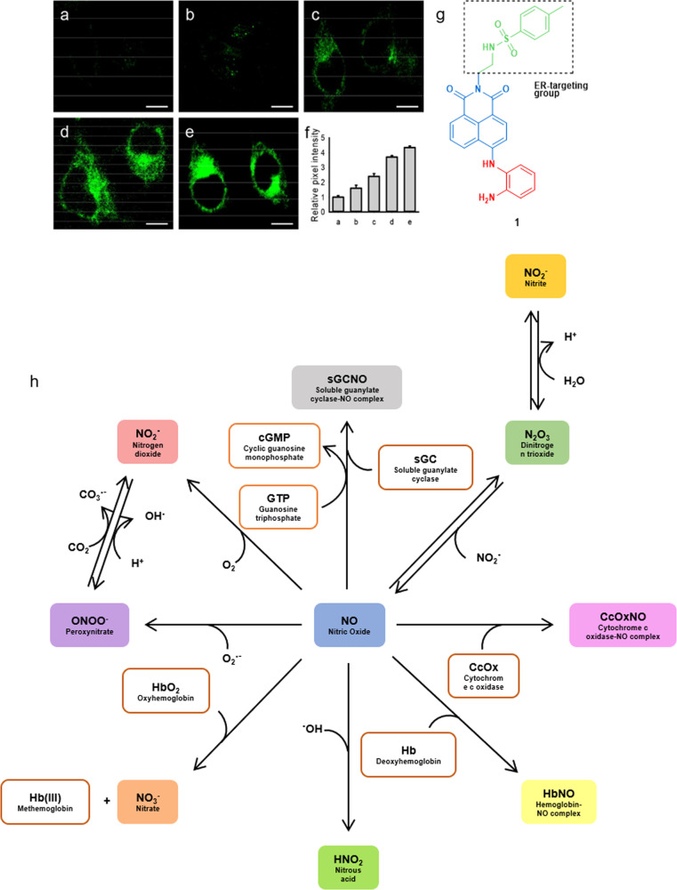
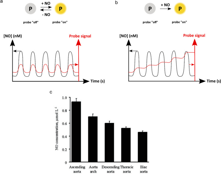
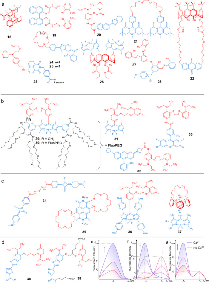
Cardiovascular diseases are the leading cause of death worldwide and are not typically diagnosed until the disease has manifested. Endothelial dysfunction is an early, reversible precursor in the irreversible development of cardiovascular diseases and is characterized by a decrease in nitric oxide production. We believe that more reliable and reproducible methods are necessary for the detection of endothelial dysfunction. Both nitric oxide and calcium play important roles in the endothelial function. Here we review different types of molecular sensors used in biological settings. Next, we review the current nitric oxide and calcium sensors available. Finally, we review methods for using both sensors for the detection of endothelial dysfunction.


Conflict of interest statement

H.E. and L.J.v.d.B. are employees of Mimetas B.V., which markets the OrganoPlate and holds the registered trademarks OrganoPlate. The other authors declare no competing interests.

Figures

Fig. 1
Fig. 1. Pros and cons of in vivo and in vitro 2D and 3D cell cultures for modelling endothelial dysfunction.
a Pros and cons of 2D culture. b Pros and cons of in vivo culture. c Pros and cons of 3D culture.
Fig. 2
Fig. 2. Nitric oxide and calcium in endothelial cells in healthy and disease setting.
a The cell signalling pathway from intracellular Ca2+ to nitric oxide in homoeostatic endothelial cell, and b in a stressed endothelial cell. c, d NO release from single endothelial cell after stimulation with Calcium Ionophore A23187 (CaI) in vitro from iliac artery of c normotensive rat and d hypotensive rat as measured with a single fibre porphyrinic sensor. eg Calcium signalling pattern in endothelial cells under resting conditions (Reprinted with permission from ref. ). e XY image of single endothelial cells showing calcium oscillations in sites 1 and 2 (marked). f Calcium concentration vs. time at the site 1. g Calcium concentration vs. time at the site 2 (Reprinted with permission from Elsevier (Burdyga et al., Copyright 2003)).
Fig. 3
Fig. 3. Targeted fluorescent imaging of endogenous NO and different pathways for biological consumption of NO.
Two-photon fluorescence images of HeLa cells treated by different concentrations of ER-stress inducer tunicamycin for 12 h, a 0, b 0.5, c 2.0, d 10.0, and e 100 μg mL−1, and then stained by the NO-sensitive molecular probe 1 (10 μM) for 20 min. Scale bar is 10 μm (Reprinted with permission from ref. . Copyright 2018 American Chemical Society). f Relative pixel intensities for images ae. Tunicamycin is a nucleoside that is commonly used to induce endoplasmic reticulum stress. g Chemical structure of the fluorescent probe 1. h Nitric oxide consumption in the biological environment.
Fig. 4
Fig. 4. Chemical structures of chosen NO sensors and example for NO sensing from endothelial cells.
a General scheme for the transformation of phenylene diamine-functionalized fluorophore to a triazole-functionalized analogue. The grey colour represents a fluorophore that emits poorly because it is quenched by PET, while the blue colour represents a fluorophore that has recovered a strong emission. b Molecular formulae of a selection of organic nitric oxide sensors based on the phenylenediamine motif,,. c General sensing method for NO by metal-based fluorescent probes. d Molecular formulae of selected examples of transition metal-based NO sensors,. e Selected nitric oxide sensors useful for sensing endothelial dysfunction. f Detection of NO produced by endothelial cells in vitro using Lippard’s molecular probe 17. (i) NO detection in porcine aortic endothelial cells (PAECs); Left: 45 min incubation of 17 (20 µM). Right: 45 min incubation of 17 (20 µM) and H2O2 (150 µM). Top: bright-field images of cells. Bottom: fluorescence images of cells. Scale bar is 50 µm. (ii) Detection of NO with 17 in Human Coronary Artery Endothelial Cells (HCAECs), with or without NO-inhibitor (L-NAME). Shown are the fluorescence images after 45 min co-incubation of the probe 17 (2 µM) with H2O2 (150 µM), L-NAME (100 µM), and/or Acetylcholine (ACh) (10 µM) according to scheme. Scale bar is 75 µm (Reprinted with permission from ref. ).
Fig. 5
Fig. 5. Schematic representation of reversible vs. irreversible NO sensor and example of NO level found in aortic endothelial cells.
a Reversible vs. b irreversible NO sensing probes, and relation of reversibility to time-dependent NO level detection. c Maximal NO concentration measured in vitro in endothelial cells of aorta (Reprinted with permission from ref. ). All NO levels measured with an electrochemical NO sensor.
Fig. 6
Fig. 6. Ca2+ intracytosolic concentration maximum in different studies.
a Resting cell characterized by low Ca2+ levels in the cell cytosol (100 nM). b Stimulated cell has increased Ca2+ levels in the cell cytosol depends on stimulus. Stimulus (Ca2+ concentration): thrombin (2 μM), acethylcholine (410 nM), shear–stress (1 μM), adenosine triphosphate (ATP) (586 μM), Brandykinin (605 nM), Hystamine (700 nM),–.
Fig. 7
Fig. 7. Chemical structures of chosen calcium sensors and graphical representation of intensometric, ratiometric and pseudo-ratiometric sensors.
a Chemical formulae of a selection of published molecular probes for calcium ions:–,–. b Calcium chemosensors based on the BAPTA chelating unit; c Calcium ions sensors that are excited by, or emit, red light–,; d Chemical formulae of a selection of ratiometric chemosensors 38, 39. eg Typical emission spectra of e an intensometric Ca2+ chemosensors, f a ratiometric Ca2+ chemosensors, and g a pseudo-ratiometric chemosensors, upon gradual addition of calcium ions.

References

    1. WHO. Cardiovascular diseases (CVDs). https://www.who.int/news-room/fact-sheets/detail/cardiovascular-diseases... (2020).
    1. Rafieian-Kopaei M, Setorki M, Doudi M, Baradaran A, Nasri H. Atherosclerosis: process, indicators, risk factors and new hopes. Int. J. Prev. Med. 2014;5:927–946. - PMC - PubMed
    1. Harvard Health Publishing. Cardiac exercise stress testing: what it can and cannot tell you. https://www.health.harvard.edu/heart-disease-overview/cardiac-exercise-s... (2020).
    1. Park K-H, Park WJ. Endothelial dysfunction: clinical implications in cardiovascular disease and therapeutic approaches. J. Korean Med. Sci. 2015;30:1213–1225. - PMC - PubMed
    1. Murad F. Discovery of some of the biological effects of nitric oxide and its role in cell signaling (Nobel Lecture) Angew. Chem. Int. Ed. 1999;38:1856–1868. - PubMed