. 2023 Nov;31(11):1251-1260.
doi: 10.1038/s41431-023-01445-2. Epub 2023 Aug 30.

CUX1-related neurodevelopmental disorder: deep insights into phenotype-genotype spectrum and underlying pathology

Affiliations

CUX1-related neurodevelopmental disorder: deep insights into phenotype-genotype spectrum and underlying pathology

Henry Oppermann et al. Eur J Hum Genet. 2023 Nov.

Abstract

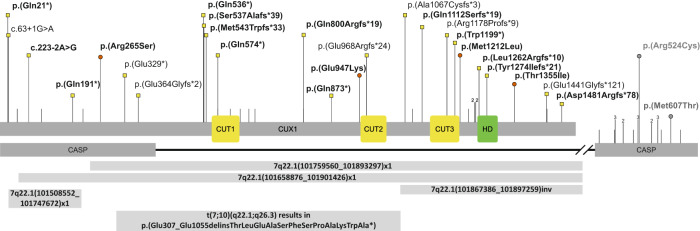
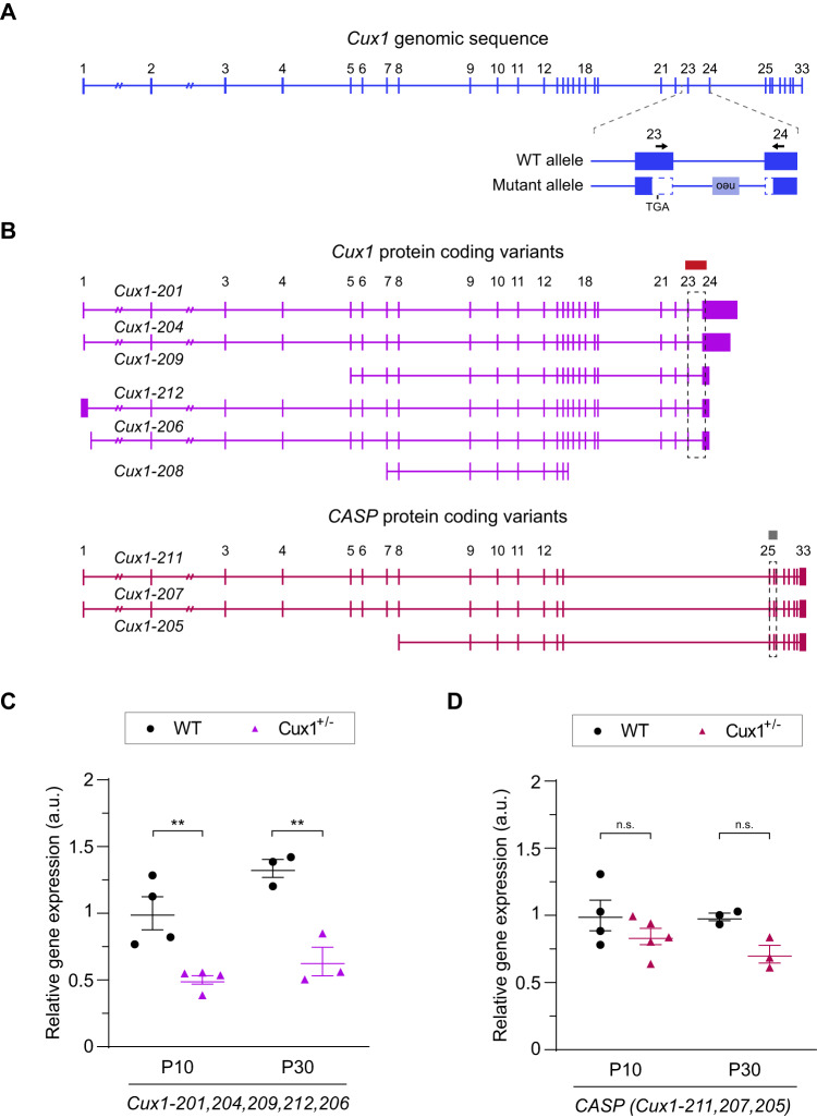
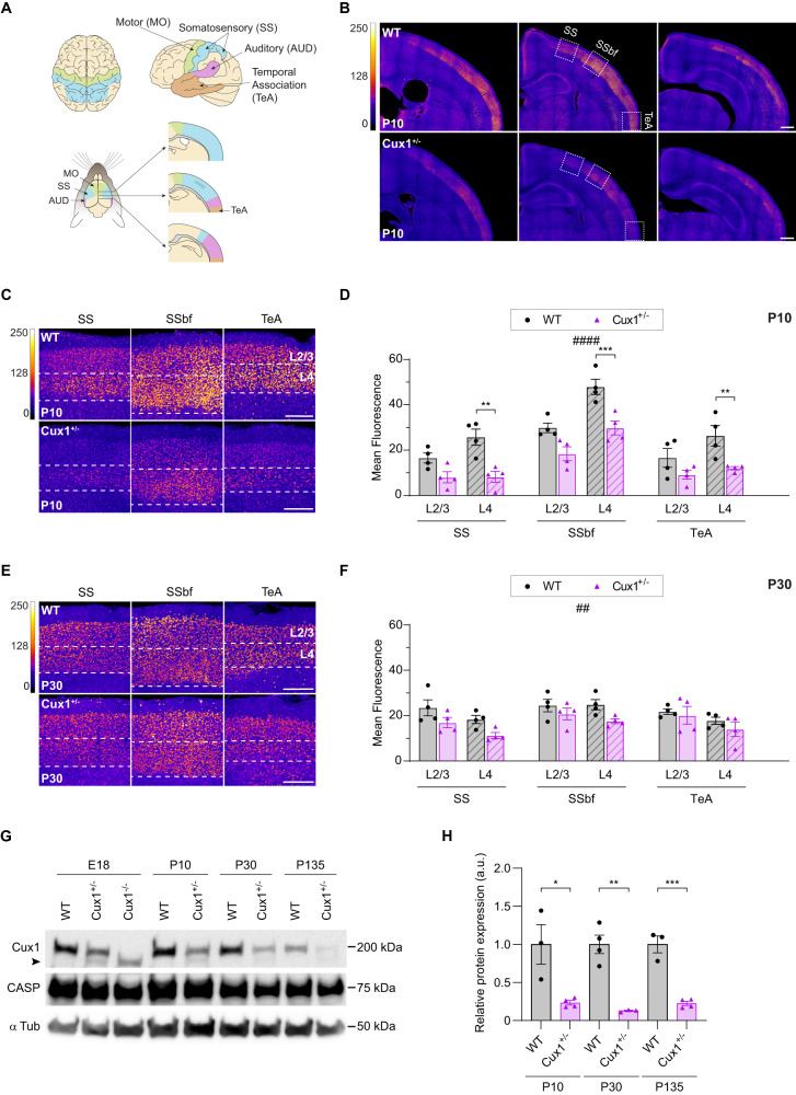
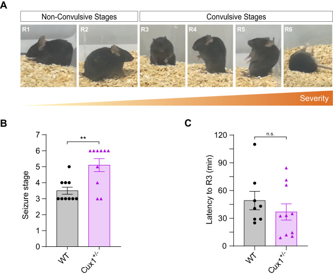
Heterozygous, pathogenic CUX1 variants are associated with global developmental delay or intellectual disability. This study delineates the clinical presentation in an extended cohort and investigates the molecular mechanism underlying the disorder in a Cux1+/- mouse model. Through international collaboration, we assembled the phenotypic and molecular information for 34 individuals (23 unpublished individuals). We analyze brain CUX1 expression and susceptibility to epilepsy in Cux1+/- mice. We describe 34 individuals, from which 30 were unrelated, with 26 different null and four missense variants. The leading symptoms were mild to moderate delayed speech and motor development and borderline to moderate intellectual disability. Additional symptoms were muscular hypotonia, seizures, joint laxity, and abnormalities of the forehead. In Cux1+/- mice, we found delayed growth, histologically normal brains, and increased susceptibility to seizures. In Cux1+/- brains, the expression of Cux1 transcripts was half of WT animals. Expression of CUX1 proteins was reduced, although in early postnatal animals significantly more than in adults. In summary, disease-causing CUX1 variants result in a non-syndromic phenotype of developmental delay and intellectual disability. In some individuals, this phenotype ameliorates with age, resulting in a clinical catch-up and normal IQ in adulthood. The post-transcriptional balance of CUX1 expression in the heterozygous brain at late developmental stages appears important for this favorable clinical course.


Conflict of interest statement

SWS is a scientific consultant for Population Bio and the King Abdullaziz University, and intellectual property held in his name by the Hospital for Sick Children is licensed to Athena Diagnostics. There is no conflicted of interest for all other authors.

Figures

Fig. 1
Fig. 1. Overview of CUX1 variants.
Location of missense and null variants in CUX1 with respect to the domain structure of CUX1 (GenBank: NM_001202543.2) and CASP (GenBank: NM_001913.4). Variants reported in this cohort are labeled with the corresponding p- or c-code and are indicated by a red circle (missense) or a yellow square (null variant). Variants that affect only CASP are labeled in gray, as the relevance of those variants is uncertain. Confirmed de novo variants are indicated in bold. Lines above the protein scheme indicate null variants in gnomAD with allele count (1 is not shown). Gross deletions and structural variants are indicated as bars below the protein scheme. Abbreviations: CUT: CUT domain, HD: CUT homeobox.
Fig. 2
Fig. 2. The levels of WT Cux1 transcripts are reduced in the cortex of Cux1+/− mice.
A Exon structure of Cux1 genomic sequence and detail showing the variant deleting exons 23 and 24 in Cux1+/−. Vertical lines represent individual exons. Arrows highlight primer sequences used for the quantifications of WT transcripts by RT-qPCR. B Predicted transcripts coding for CUX1 and CASP. Boxes and dashed boxes highlight the regions containing the RT-qPCR amplicons used to quantify protein-coding transcripts. The Cux1 amplicon measures all annotated CUX1 protein-coding transcripts (Cux1-201, 204, 209, 212, 206) except Cux1-208. RT-qPCR amplicon for CASP measures all annotated CASP protein-coding transcripts (Cux1-211, 207, 205). C Relative expression of Cux1 protein-coding transcript isoforms (Cux1-201, 204, 209, 212, 206) as shown in (A) and (B) at P10 and P30, quantified by RT-q-PCR. Data are shown normalized to P10 WT levels. Data show mean ± SEM (n ≥ 3 animals per condition. Two-way ANOVA: P-value WT vs. Cux+/- #### ≤0.0001. Post hoc with Tukey´s test: P-value P10 WT vs. Cux1+/− ** ≤0.005, P-value P30 WT vs. Cux+/−** ≤0.005). D Relative gene expression of protein-coding CASP transcripts at P10 and P30. Data are shown normalized to P10 WT levels. Data show mean ± SEM (n ≥ 3 animals per condition. Two-way ANOVA: P-value WT vs. Cux+/- # ≤0.05. Post hoc with Tukey´s test: P-value P10 WT vs. Cux+/−  = 0.4641 (n.s.), P-value P30 WT vs. Cux+/−  = 0.1846 (n.s.)).
Fig. 3
Fig. 3. Cux1 cortical expression is reduced in heterozygous mice.
A Comparative scheme of distinct functional areas in human (top) and mouse (bottom) brains. Top, dorsal (left), and lateral (right) views of motor (MO, green), somatosensory (SS, blue), auditory (AUD, magenta), and temporal association (TeA) cortical areas in the human brain. Bottom, dorsal (left), and medio-lateral views at several anteroposterior coordinates (right) of the functional areas in the mouse brain. B Intensity maps of Cux1 expression early in development (P10) in WT and Cux1+/− mouse brains. Images show coronal sections from more anterior (left) to more posterior (right) coordinates. Dashed boxes in the middle images highlight areas of interest (SSbf, somatosensory barrel field). Scale bar = 500 µm. C, E Magnified images of cortical upper-layer neurons (L2-4) from dashed areas of Fig. S1B at P10 (C) and P30 (E). Scale bar = 200 µm. D, F Quantification of Cux1 expression in upper-layer neurons of SS, SSbf, and TeA areas at P10 (D) and P30 (F). Data show mean ± SEM (n ≥ 3 animals per condition, n = 2 sections per brain. P10 Two-way ANOVA: P-value WT vs. Cux1+/− #### ≤ 0.0001. Post hoc with Sidak´s test: P-value SSL4 WT vs. Cux1+/− ** ≤ 0.01, P-value SSbfL4 WT vs. Cux+/− *** ≤ 0.001, P-value TeAL4 WT vs. Cux+/− ** ≤ 0.01. P30 Two-way ANOVA: P-value WT vs. Cux1+/−## ≤ 0.01.). G Western blot showing cortical expression of the full-length 200 kDa CUX1 in WT, Cux1+/−, and Cux1/− (E18 only) mice at E18, P10, P30, and P135. The amount of protein was quantified and normalized to α tubulin expression (50 kDa). CASP (75 kDa), an alternatively spliced product of the Cux1 gene, is also recognized by this antibody. The truncated mutant CUX1 is indicated by an arrowhead. H Relative cortical expression of the 200 kDa CUX1 at P10, P30, and P135. Data show mean ± SEM. (n = 3–4 cortical samples per condition. P10 unpaired t test: P-value WT vs. Cux1+/− * ≤ 0.05. P30 unpaired t test: P-value WT vs. Cux1+/− ** ≤ 0.01. P135 unpaired t test: P-value WT vs. Cux+/− *** ≤ 0.001).
Fig. 4
Fig. 4. Cux1 heterozygosity results in increased seizure susceptibility in mice.
A Identification of stages of seizure in a kainate-induced model of epilepsy. Photographs show representative behaviors from the less severe to the most, progressing from non-convulsive to convulsive seizure stages (R1-R6). B The maximum stage of seizure was reached in WT and Cux1+/− mice during the first 2 h of kainate induction. Data show mean ± SEM (n = 10 per condition. Mann–Whitney test: P-value WT vs. Cux1+/− ** ≤ 0.01). C Latency to the onset of convulsive stages (R3) in WT and Cux1+/− mice after kainate induction. Data show mean ± SEM (n ≥ 8 per condition. Mann–Whitney test: P-value WT vs. Cux+/− = 0.3478 (n.s.)).

References

    1. Sansregret L, Nepveu A. The multiple roles of CUX1: insights from mouse models and cell-based assays. Gene. 2008;412:84–94. doi: 10.1016/j.gene.2008.01.017. - DOI - PubMed
    1. Gillingham AK, Pfeifer AC, Munro S. CASP, the alternatively spliced product of the gene encoding the CCAAT-displacement protein transcription factor, Is a Golgi membrane protein related to giantin. Mol Biol Cell. 2002;13:3761–74. doi: 10.1091/mbc.e02-06-0349. - DOI - PMC - PubMed
    1. Hulea L, Nepveu A. CUX1 transcription factors: from biochemical activities and cell-based assays to mouse models and human diseases. Gene. 2012;497:18–26. - PubMed
    1. Weiss LA, Nieto M. The crux of Cux genes in neuronal function and plasticity. Brain Res. 2019;1705:32–42. doi: 10.1016/j.brainres.2018.02.044. - DOI - PubMed
    1. Saito T, Hanai S, Takashima S, Nakagawa E, Okazaki S, Inoue T, et al. Neocortical layer formation of human developing brains and lissencephalies: consideration of layer-specific marker expression. Cereb Cortex. 2011;21:588–96. doi: 10.1093/cercor/bhq125. - DOI - PubMed

Publication types