

Ocular manifestations of renal ciliopathies
- PMID: 37644229
- PMCID: PMC10942941
- DOI: 10.1007/s00467-023-06096-5
Ocular manifestations of renal ciliopathies
Abstract
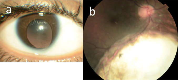
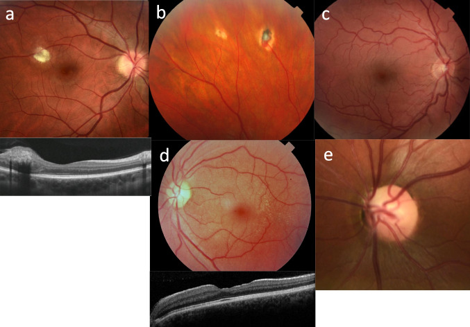
Renal ciliopathies are a common cause of kidney failure in children and adults, and this study reviewed their ocular associations. Genes affected in renal ciliopathies were identified from the Genomics England Panels. Ocular associations were identified from Medline and OMIM, and the genes additionally examined for expression in the human retina ( https://www.proteinatlas.org/humanproteome/tissue ) and for an ocular phenotype in mouse models ( http://www.informatics.jax.org/ ). Eighty-two of the 86 pediatric-onset renal ciliopathies (95%) have an ocular phenotype, including inherited retinal degeneration, oculomotor disorders, and coloboma. Diseases associated with pathogenic variants in ANK6, MAPKBP1, NEK8, and TCTN1 have no reported ocular manifestations, as well as low retinal expression and no ocular features in mouse models. Ocular abnormalities are not associated with the most common adult-onset "cystic" kidney diseases, namely, autosomal dominant (AD) polycystic kidney disease and the AD tubulointerstitial kidney diseases (ADTKD). However, other kidney syndromes with cysts have ocular features including papillorenal syndrome (optic disc dysplasia), Hereditary Angiopathy Nephropathy, Aneurysms and muscle Cramps (HANAC) (tortuous retinal vessels), tuberous sclerosis (retinal hamartomas), von Hippel-Lindau syndrome (retinal hemangiomas), and Alport syndrome (lenticonus, fleck retinopathy). Ocular abnormalities are associated with many pediatric-onset renal ciliopathies but are uncommon in adult-onset cystic kidney disease. However the demonstration of ocular manifestations may be helpful diagnostically and the features may require monitoring or treatment.
Keywords: Coloboma; Inherited retinal degeneration; Nephronophthisis; Oculomotor disorder; Renal ciliopathy.
© 2023. The Author(s).
Conflict of interest statement
OS was a medical student who undertook this research project as part of his degree. None of the authors has a financial or non-financial Conflict of Interest to declare.
Figures

Similar articles
-
Alport syndrome. A review of the ocular manifestations.Ophthalmic Genet. 1997 Dec;18(4):161-73. doi: 10.3109/13816819709041431. Ophthalmic Genet. 1997. PMID: 9457747 Review.
-
Phenotypic Spectrum of Children with Nephronophthisis and Related Ciliopathies.Clin J Am Soc Nephrol. 2017 Dec 7;12(12):1974-1983. doi: 10.2215/CJN.01280217. Epub 2017 Nov 16. Clin J Am Soc Nephrol. 2017. PMID: 29146700 Free PMC article.
-
Ocular features in Alport syndrome: pathogenesis and clinical significance.Clin J Am Soc Nephrol. 2015 Apr 7;10(4):703-9. doi: 10.2215/CJN.10581014. Epub 2015 Feb 3. Clin J Am Soc Nephrol. 2015. PMID: 25649157 Free PMC article. Review.
-
The retinal "lozenge" or "dull macular reflex" in Alport syndrome may be associated with a severe retinopathy and early-onset renal failure.Br J Ophthalmol. 2009 Mar;93(3):383-6. doi: 10.1136/bjo.2008.142869. Epub 2008 Nov 19. Br J Ophthalmol. 2009. PMID: 19019929
-
Repurposing small molecules for nephronophthisis and related renal ciliopathies.Kidney Int. 2023 Aug;104(2):245-253. doi: 10.1016/j.kint.2023.04.027. Epub 2023 May 25. Kidney Int. 2023. PMID: 37244473 Review.
Cited by
-
Extrarenal Clinical Features are Reported for Most Genes Implicated in Genetic Kidney Disease.Kidney Int Rep. 2025 Feb 5;10(4):1196-1204. doi: 10.1016/j.ekir.2025.01.045. eCollection 2025 Apr. Kidney Int Rep. 2025. PMID: 40303230 Free PMC article.
-
A Novel NPHP5 Gene Mutation in Three Siblings With Nephronophthisis Without Retinitis Pigmentosa: A Case Report.Case Rep Genet. 2025 Apr 28;2025:1453255. doi: 10.1155/crig/1453255. eCollection 2025. Case Rep Genet. 2025. PMID: 40331022 Free PMC article.
References
Publication types
MeSH terms
LinkOut - more resources
Full Text Sources
Medical
