

A mini-invasive surgical technique for Carlevale IOL implantation: case series study and description of concomitant surgery
- PMID: 37644329
- PMCID: PMC10844417
- DOI: 10.1007/s00417-023-06217-8
A mini-invasive surgical technique for Carlevale IOL implantation: case series study and description of concomitant surgery
Abstract
Purpose: To examine the feasibility and outcomes of a modified technique for the implantation of scleral fixated Carlevale intraocular lens (IOL) (I71 FIL SSF. Soleko IOL Division, Pontecorvo, Italy), and to analyze the occurrence of adverse events.
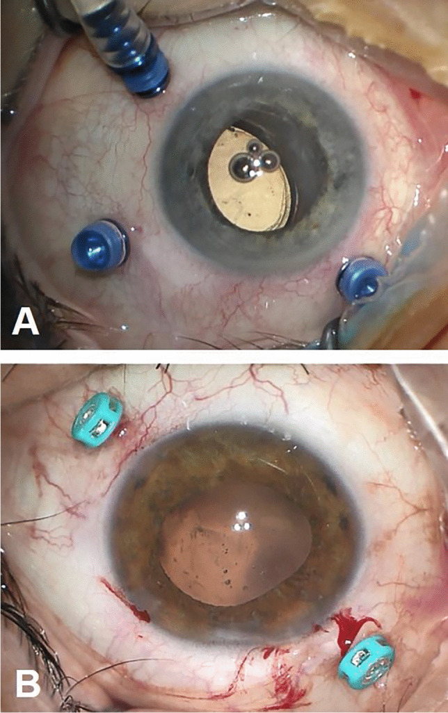
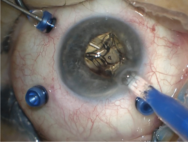
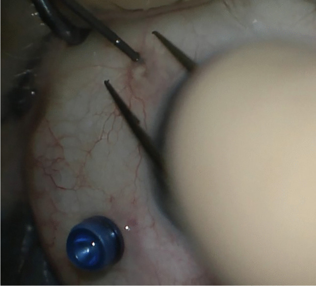
Methods: This is a retrospective observational study conducted revising patients charts from 2018 to 2023. Thirty-five eyes of 33 patients were included. Patients requiring IOL explantation had either IOL dislocation or opacification. The implantation of the Carlevale IOL was performed with the subconjunctival positioning of the anchors without any scleral flap. All maneuvers were performed transconjunctivally. The anatomical outcomes considered were IOL positioning, and the absence of postoperative complications. The functional outcomes analyzed were best correctedvisual acuity (BCVA) and refraction.
Results: In all the cases, the IOL was well positioned and centered postoperatively. No cases of conjunctival erosion were recorded. The best corrected visual acuity (BCVA) was 0.9±0.6 logMar (mean±standard deviation) preoperatively and 0.5±0.5 logMar (mean±standard deviation) postoperatively. The mean preoperative spherical equivalent was +6.8±7.7 dioptres, while postoperatively it was -1.1±1.6 dioptres. The most frequent procedure associated to secondary IOL implantation was posterior vitrectomy (25 eyes, 71.4%), which was performed with 25-gauge transconjunctival cannulas in the ciliary sulcus. The follow-up period was 24.5±16.9 months (mean±standard deviation).
Conclusion: The described mini-invasive technique for Carlevale IOL implantation is safe and effective. It can be recommended either as a stand-alone operation or associated to concurrent surgical procedures.
Keywords: Carlevale IOL; IOL luxation; IOL opacification; Scleral fixation; Secondary IOL implantation.
© 2023. The Author(s).
Conflict of interest statement
Carla Danese is consultant for Bayer, outside the submitted work. Francesco Di Bin declares no conflicts of interest. Paolo Lanzetta is consultant for Aerie, Allergan, Apellis, Bausch&Lomb, Bayer, Biogen, Boerhinger, Centervue, Genentech, Novartis, Ocular Therapeutix, Outlook Therapeutics, Roche, outside the submitted work.
Figures


Similar articles
-
FIL-SSF Carlevale intraocular lens for sutureless scleral fixation: 7 recommendations from a serie of 72 cases. MICA study (Multicentric Study of the Carlevale IOL).J Fr Ophtalmol. 2021 Sep;44(7):1038-1046. doi: 10.1016/j.jfo.2021.05.002. Epub 2021 Jun 17. J Fr Ophtalmol. 2021. PMID: 34148705
-
Comparison of two different scleral fixation techniques of posterior chamber Carlevale lens.Medicine (Baltimore). 2021 Aug 13;100(32):e26728. doi: 10.1097/MD.0000000000026728. Medicine (Baltimore). 2021. PMID: 34397876 Free PMC article.
-
Refractive outcomes for secondary sutureless posterior chamber lens implantation: sutureless scleral fixating lens Carlevale® versus retropupillary iris-claw lens Artisan®.Graefes Arch Clin Exp Ophthalmol. 2025 Mar;263(3):735-743. doi: 10.1007/s00417-024-06683-8. Epub 2024 Nov 11. Graefes Arch Clin Exp Ophthalmol. 2025. PMID: 39527243 Free PMC article.
-
SCLERAL FIXATION OF CARLEVALE INTRAOCULAR LENS: A Systematic Review and Meta-Analysis.Retina. 2023 Oct 1;43(10):1750-1762. doi: 10.1097/IAE.0000000000003873. Retina. 2023. PMID: 37399540
-
FIL SSF intraocular lens opacification after pars plana vitrectomy with gas tamponade for traumatic lens luxation and retinal detachment: a case report and literature review.BMC Ophthalmol. 2023 Sep 25;23(1):383. doi: 10.1186/s12886-023-03126-6. BMC Ophthalmol. 2023. PMID: 37743488 Free PMC article. Review.
Cited by
-
Recent advances and current challenges in suture and sutureless scleral fixation techniques for intraocular lens: a comprehensive review.Eye Vis (Lond). 2024 Dec 30;11(1):49. doi: 10.1186/s40662-024-00414-0. Eye Vis (Lond). 2024. PMID: 39736769 Free PMC article. Review.
-
Current Evidence for a New Surgical Technique for Scleral Fixation: The Implantation of a Carlevale Lens, a Systematic Review.J Clin Med. 2024 Jun 3;13(11):3287. doi: 10.3390/jcm13113287. J Clin Med. 2024. PMID: 38892997 Free PMC article. Review.
-
Long-term results after sutureless intrascleral fixation of the Carlevale intraocular lens: Changes in scleral pocket thickness over time.Acta Ophthalmol. 2025 Aug;103(5):e290-e297. doi: 10.1111/aos.17468. Epub 2025 Feb 27. Acta Ophthalmol. 2025. PMID: 40013529 Free PMC article. Clinical Trial.
-
Implantation of Sutureless Scleral-Fixated Carlevale Intraocular Lens (IOL) in Patients with Insufficient Capsular Bag Support: A Retrospective Analysis of 100 Cases at a Single Center.J Clin Med. 2025 Jun 19;14(12):4378. doi: 10.3390/jcm14124378. J Clin Med. 2025. PMID: 40566120 Free PMC article.
References
-
- Czajka MP, Frajdenberg A, Stopa M, Pabin T, Johansson B, Jakobsson G. Sutureless intrascleral fixation using different three- piece posterior chamber intraocular lenses: a literature review of surgical techniques in cases of insufficient capsular support and a retrospective multicentre study. Acta Ophthalmol. 2020;98:224–236. doi: 10.1111/aos.14307. - DOI - PubMed
Publication types
MeSH terms
LinkOut - more resources
Full Text Sources
