

Diagnosis of thoracic outlet syndrome with the lower trunk compression of brachial plexus by high-frequency ultrasonography
- PMID: 37644436
- PMCID: PMC10463735
- DOI: 10.1186/s12891-023-06762-7
Diagnosis of thoracic outlet syndrome with the lower trunk compression of brachial plexus by high-frequency ultrasonography
Abstract
Background: Thoracic outlet syndrome (TOS) with the lower trunk compression of brachial plexus (BP) is difficult to diagnosis. This study aimed to summarize the features of thoracic outlet syndrome (TOS) with the lower trunk compression of brachial plexus observed on high-frequency ultrasonography (HFUS).
Methods: The ultrasound data of 27 patients who had TOS with the lower trunk compression of brachial plexus were collected and eventually confirmed by surgery. The imaging data were compared, and the pathogenesis of TOS was analyzed on the basis of surgical data.
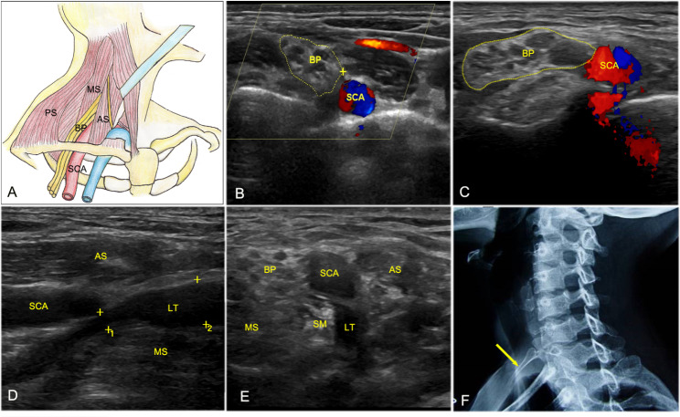
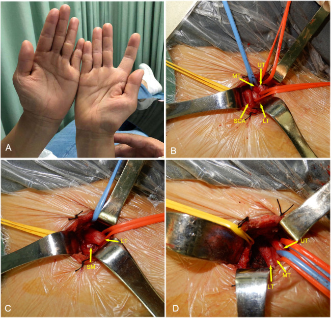
Results: TOS occurred predominantly in females (70.4%). Most cases had unilateral involvement (92.6%), mainly on the right side (66.7%). The HFUS features of TOS can be summarized as follows: (1) Lower trunk compression. HFUS revealed focal thinning that reflected compression at the level of the lower trunk; furthermore, the distal part of the nerve was thickened for edema (Affected side: 0.49 ± 0.12 cm vs. Healthy side: 0.38 ± 0.06, P = 0.009), and the cross-sectional area of brachial plexus cords was markedly greater on the injured side than on the healthy side (0.95 ± 0.08 cm² vs. 0.65 ± 0.11 cm², P = 0.004). (2) Hyperechoic fibromuscular bands behind the compressed nerve (mostly the scalenus minimus muscle). (3) Abnormal bony structures: cervical ribs or elongated transverse processes of the 7th cervical vertebra (C7). Surgical results showed that the etiological factors contributing to TOS were (1) muscle hypertrophy and/or fibrosis (100%) and (2) cervical ribs/elongated C7 transverse processes (20.7%).
Conclusion: TOS with the lower trunk compression of brachial plexus can be diagnosed accurately and reliably by high-frequency ultrasound.
Keywords: Brachial plexus; High-frequency ultrasonography; Thoracic outlet syndrome.
© 2023. BioMed Central Ltd., part of Springer Nature.
Conflict of interest statement
The authors declare no competing interests.
Figures
References
-
- Le Forestier N, Mouton P, Maisonobe T, et al. True neurological thoracic outlet syndrome. Rev Neurol (Paris) 2000;156(1):34–40. - PubMed
MeSH terms
Grants and funding
LinkOut - more resources
Full Text Sources
Medical
Miscellaneous
