

Elevated expression of the RNA-binding protein IGF2BP1 enhances the mRNA stability of INHBA to promote the invasion and migration of esophageal squamous cancer cells
- PMID: 37644505
- PMCID: PMC10466848
- DOI: 10.1186/s40164-023-00429-8
Elevated expression of the RNA-binding protein IGF2BP1 enhances the mRNA stability of INHBA to promote the invasion and migration of esophageal squamous cancer cells
Abstract
Background: The mechanisms underlying the occurrence and development of esophageal squamous cell carcinoma (ESCC) remains to be elucidated. The present study aims to investigate the roles and implications of IGF2BP1 overexpression in ESCC.
Methods: IGF2BP1 protein expression in ESCC samples was assessed by immunohistochemistry (IHC), and the mRNA abundance of IGF2BP1 and INHBA was analyzed with TCGA datasets and by RNA in situ hybridization (RISH). The methylation level of the IGF2BP1 promoter region was detected by methylation-specific PCR (MSP-PCR). Cell viability, migration, invasion and in vivo metastasis assays were performed to explore the roles of IGF2BP1 overexpression in ESCC. RNA immunoprecipitation sequencing (RIP-seq) and mass spectrometry were applied to identify the target RNAs and interacting proteins of IGF2BP1, respectively. RIP-PCR, RNA pulldown, immunofluorescence (IF), gene-specific m6A PCR and RNA stability assays were used to uncover the molecular mechanisms underlying the malignant phenotypes of ESCC cells caused by IGF2BP1 dysregulation. BTYNB, a small molecular inhibitor of IGF2BP1, was evaluated for its inhibitory effect on the malignant phenotypes of ESCC cells.
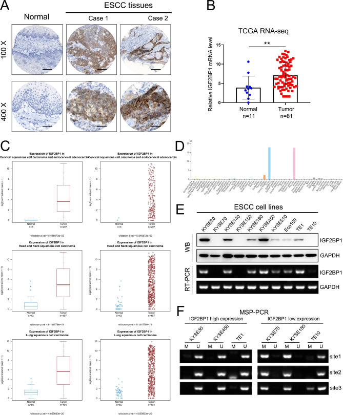
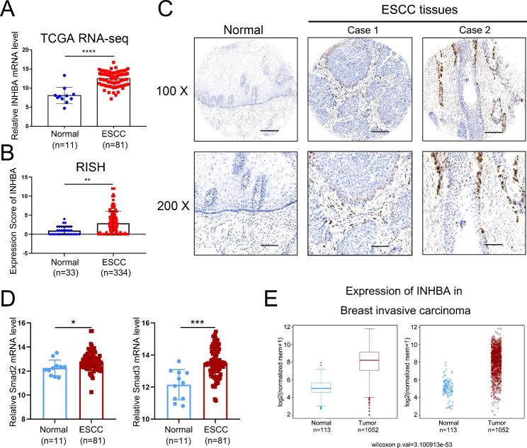
Results: IGF2BP1 overexpression was detected in ESCC tissues and associated with the depth of tumor invasion. In addition, IGF2BP1 mRNA expression in ESCC cells was negatively correlated with the level of its promoter methylation. Knockdown of IGF2BP1 inhibited ESCC cell invasion and migration as well as tumor metastasis. Mechanistically, we observed that IGF2BP1 bound and stabilized INHBA mRNA and then resulted in higher protein expression of INHBA, leading to the activation of Smad2/3 signaling, thus promoting malignant phenotypes. The mRNA level of INHBA was upregulated in ESCC tissues as well. Furthermore, IGF2BP1 interacted with G3BP stress granule assembly factor 1 (G3BP1). Knockdown of G3BP1 also down-regulated the INHBA-Smad2/3 signaling. BTYNB abolished this activated signaling and significantly attenuated the malignant phenotypes of ESCC cells.
Conclusions: Elevated expression of IGF2BP1 is a frequent event in ESCC tissues and might be a candidate biomarker for the disease. IGF2BP1 overexpression promotes the invasion and migration of ESCC cells by activating the INHBA-Smad2/3 pathway, providing a potential therapeutic target for ESCC patients with high expression of IGF2BP1.
Keywords: Esophageal squamous cell carcinoma; IGF2BP1; INHBA; Invasion; Migration; RNA binding protein.
© 2023. YUMED Inc. and BioMed Central Ltd.
Conflict of interest statement
The authors declare no competing interests.
Figures


Similar articles
-
Insulin-like growth factor 2 mRNA-binding protein 1 (IGF2BP1) in hematological diseases.Mol Med. 2024 Sep 28;30(1):165. doi: 10.1186/s10020-024-00936-2. Mol Med. 2024. PMID: 39342091 Free PMC article. Review.
-
PABPC1-induced stabilization of IFI27 mRNA promotes angiogenesis and malignant progression in esophageal squamous cell carcinoma through exosomal miRNA-21-5p.J Exp Clin Cancer Res. 2022 Mar 28;41(1):111. doi: 10.1186/s13046-022-02339-9. J Exp Clin Cancer Res. 2022. PMID: 35346324 Free PMC article.
-
Circular RNA circNTRK2 facilitates the progression of esophageal squamous cell carcinoma through up-regulating NRIP1 expression via miR-140-3p.J Exp Clin Cancer Res. 2020 Jul 11;39(1):133. doi: 10.1186/s13046-020-01640-9. J Exp Clin Cancer Res. 2020. PMID: 32653032 Free PMC article.
-
IGF2BP1 enhances the stability of SIK2 mRNA through m6A modification to promote non-small cell lung cancer progression.Biochem Biophys Res Commun. 2023 Dec 3;684:149113. doi: 10.1016/j.bbrc.2023.10.045. Epub 2023 Oct 12. Biochem Biophys Res Commun. 2023. PMID: 37866243
-
NEFL promotes invasion and migration of esophageal squamous carcinoma cells via the EGFR/AKT/S6 pathway.Yi Chuan. 2022 Apr 20;44(4):322-334. doi: 10.16288/j.yczz.22-019. Yi Chuan. 2022. PMID: 35437240
Cited by
-
Positive GLI1/INHBA feedback loop drives tumor progression in gastric cancer.Cancer Sci. 2024 Jul;115(7):2301-2317. doi: 10.1111/cas.16193. Epub 2024 Apr 27. Cancer Sci. 2024. PMID: 38676428 Free PMC article.
-
The m6A regulators in prostate cancer: molecular basis and clinical perspective.Front Pharmacol. 2024 Aug 29;15:1448872. doi: 10.3389/fphar.2024.1448872. eCollection 2024. Front Pharmacol. 2024. PMID: 39268470 Free PMC article. Review.
-
CircRNF13 enhances IGF2BP1 phase separation-mediated ITGB1 mRNA stabilization in an m6A-dependent manner to promote oral cancer cisplatin chemoresistance.Mol Cancer. 2025 Jan 31;24(1):36. doi: 10.1186/s12943-025-02239-4. Mol Cancer. 2025. PMID: 39891203 Free PMC article.
-
The Biological Role of LRPPRC in Human Cancers.Cancer Control. 2025 Jan-Dec;32:10732748251353077. doi: 10.1177/10732748251353077. Epub 2025 Jun 30. Cancer Control. 2025. PMID: 40587247 Free PMC article. Review.
-
Insulin-like growth factor 2 mRNA-binding protein 1 (IGF2BP1) in hematological diseases.Mol Med. 2024 Sep 28;30(1):165. doi: 10.1186/s10020-024-00936-2. Mol Med. 2024. PMID: 39342091 Free PMC article. Review.
References
-
- Zeng H, Chen W, Zheng R, Zhang S, Ji JS, Zou X, et al. Changing cancer survival in China during 2003-15: a pooled analysis of 17 population-based cancer registries. Lancet Glob Health. 2018;6(5):e555–e67. - PubMed
-
- Elcheva I, Tarapore RS, Bhatia N, Spiegelman VS. Overexpression of mRNA-binding protein CRD-BP in malignant melanomas. Oncogene. 2008;27(37):5069–74. - PubMed
-
- Kobel M, Weidensdorfer D, Reinke C, Lederer M, Schmitt WD, Zeng K, et al. Expression of the RNA-binding protein IMP1 correlates with poor prognosis in ovarian carcinoma. Oncogene. 2007;26(54):7584–9. - PubMed
Grants and funding
LinkOut - more resources
Full Text Sources
Research Materials
Miscellaneous
