

Knockdown of TFRC suppressed the progression of nasopharyngeal carcinoma by downregulating the PI3K/Akt/mTOR pathway
- PMID: 37644594
- PMCID: PMC10466839
- DOI: 10.1186/s12935-023-02995-7
Knockdown of TFRC suppressed the progression of nasopharyngeal carcinoma by downregulating the PI3K/Akt/mTOR pathway
Abstract
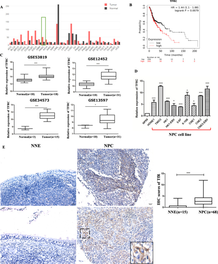
Background: The transferrin receptor (TfR) encoded by TFRC gene is the main cellular iron importer. TfR is highly expressed in many cancers and is expected to be a promising new target for cancer therapy; however, its role in nasopharyngeal carcinoma (NPC) remains unknown.
Methods: The TfR levels were investigated in NPC tissues and cell lines using immunohistochemistry and reverse transcription-quantitative polymerase chain reaction. Knockdown of TFRC using two siRNA to investigate the effects on intracellular iron level and biological functions, including proliferation by CKK-8 assay, colony formation, cell apoptosis and cell cycle by flow cytometry, migration and invasion, and tumor growth in vivo by nude mouse xenografts. RNA sequencing was performed to find possible mechanism after TFRC knockdown on NPC cells and further verified by western blotting.
Results: TfR was overexpressed in NPC cell lines and tissues. Knockdown of TFRC inhibited cell proliferation concomitant with increased apoptosis and cell cycle arrest, and it decreased intracellular iron, colony formation, migration, invasion, and epithelial-mesenchymal transition in HK1-EBV cells. Western blotting showed that TFRC knockdown suppressed the levels of the iron storage protein FTH1, anti-apoptotic marker BCL-xL, and epithelial-mesenchymal transition markers. We confirmed in vivo that TFRC knockdown also inhibited NPC tumor growth and decreased Ki67 expression in tumor tissues of nude mouse xenografts. RNA sequencing and western blotting revealed that TFRC silencing inhibited the PI3K/Akt/mTOR signaling pathway.
Conclusions: These results indicated that TfR was overexpressed in NPC, and TFRC knockdown inhibited NPC progression by suppressing the PI3K/Akt/mTOR signaling pathway. Thus, TfR may serve as a novel biomarker and therapeutic target for NPC.
Keywords: Nasopharyngeal carcinoma; PI3K/Akt/mTOR signaling pathway; RNA-seq; TFRC; siRNA.
© 2023. BioMed Central Ltd., part of Springer Nature.
Conflict of interest statement
The authors declare no competing interests.
Figures


Similar articles
-
C2orf40 inhibits metastasis and regulates chemo-resistance and radio-resistance of nasopharyngeal carcinoma cells by influencing cell cycle and activating the PI3K/AKT/mTOR signaling pathway.J Transl Med. 2022 Jun 8;20(1):264. doi: 10.1186/s12967-022-03446-z. J Transl Med. 2022. PMID: 35676661 Free PMC article.
-
Pre-treatment with angiotensin-(1-7) inhibits tumor growth via autophagy by downregulating PI3K/Akt/mTOR signaling in human nasopharyngeal carcinoma xenografts.J Mol Med (Berl). 2018 Dec;96(12):1407-1418. doi: 10.1007/s00109-018-1704-z. Epub 2018 Oct 29. J Mol Med (Berl). 2018. PMID: 30374682 Free PMC article.
-
Silencing of NACC1 inhibits the proliferation, migration and invasion of nasopharyngeal carcinoma cells via regulating the AKT/mTOR signaling pathway.Oncol Lett. 2021 Dec;22(6):828. doi: 10.3892/ol.2021.13088. Epub 2021 Oct 12. Oncol Lett. 2021. PMID: 34691255 Free PMC article.
-
Aloe-emodin mediates the inhibitory effect of LncRNA D63785 on the PI3K/Akt/mTOR pathway in nasopharyngeal carcinoma.Front Pharmacol. 2025 Jul 18;16:1573408. doi: 10.3389/fphar.2025.1573408. eCollection 2025. Front Pharmacol. 2025. PMID: 40756984 Free PMC article.
-
Targeting Glutamine Metabolism through Glutaminase Inhibition Suppresses Cell Proliferation and Progression in Nasopharyngeal Carcinoma.Anticancer Agents Med Chem. 2023;23(17):1944-1957. doi: 10.2174/1871520623666230727104825. Anticancer Agents Med Chem. 2023. PMID: 37497684
Cited by
-
Nanoscale strategies: doxorubicin resistance challenges and enhancing cancer therapy with advanced nanotechnological approaches.Drug Deliv Transl Res. 2025 Feb 15. doi: 10.1007/s13346-025-01790-3. Online ahead of print. Drug Deliv Transl Res. 2025. PMID: 39955406
-
The TFRC as a prognostic biomarker and potential therapeutic target in cervical cancer: a preliminary study.Front Oncol. 2025 Apr 15;15:1523137. doi: 10.3389/fonc.2025.1523137. eCollection 2025. Front Oncol. 2025. PMID: 40303995 Free PMC article.
-
Synthesis and Evaluation of [64Cu]Cu-NOTA-HFn for PET Imaging of Transferrin Receptor 1 Expression in Nasopharyngeal Carcinoma.ACS Omega. 2024 Apr 5;9(15):17423-17431. doi: 10.1021/acsomega.4c00187. eCollection 2024 Apr 16. ACS Omega. 2024. PMID: 38645324 Free PMC article.
-
Prolactin Drives Iron Release from Macrophages and Uptake in Mammary Cancer Cells through CD44.Int J Mol Sci. 2024 Aug 16;25(16):8941. doi: 10.3390/ijms25168941. Int J Mol Sci. 2024. PMID: 39201626 Free PMC article.
-
Carboxylesterase 4A Inhibits the Malignant Biological Behavior of Nasopharyngeal Carcinoma via the PI3K/AKT Pathway.Technol Cancer Res Treat. 2025 Jan-Dec;24:15330338251319144. doi: 10.1177/15330338251319144. Technol Cancer Res Treat. 2025. PMID: 39912257 Free PMC article.
References
Grants and funding
LinkOut - more resources
Full Text Sources
Other Literature Sources
Research Materials
Miscellaneous
