. 2023 Aug 14:11:1247638.
doi: 10.3389/fped.2023.1247638. eCollection 2023.

Ultrastructure for the diagnosis of primary ciliary dyskinesia in South Africa, a resource-limited setting

Affiliations

Ultrastructure for the diagnosis of primary ciliary dyskinesia in South Africa, a resource-limited setting

Monica Birkhead et al. Front Pediatr. .

Abstract

Introduction: International guidelines recommend a multi-faceted approach for successful diagnoses of primary ciliary dyskinesia (PCD). In the absence of a gold standard test, a combination of genetic testing/microscopic analysis of structure and function/nasal nitric oxide measurement is used. In resource-limited settings, often none of the above tests are available, and in South Africa, only transmission electron microscopy (TEM) is available in central anatomical pathology departments. The aim of this study was to describe the clinical and ultrastructural findings of suspected PCD cases managed by pediatric pulmonologists at a tertiary-level state funded hospital in Johannesburg.

Methods: Nasal brushings were taken from 14 children with chronic respiratory symptoms in keeping with a PCD phenotype. Ultrastructural analysis in accordance with the international consensus guidelines for TEM-PCD diagnostic reporting was undertaken.

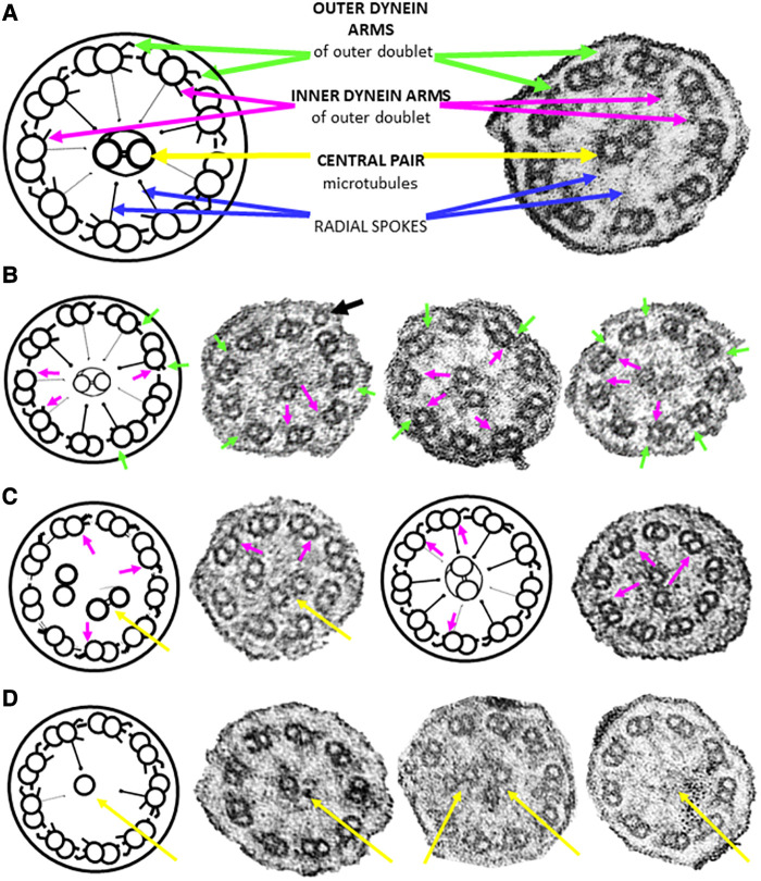
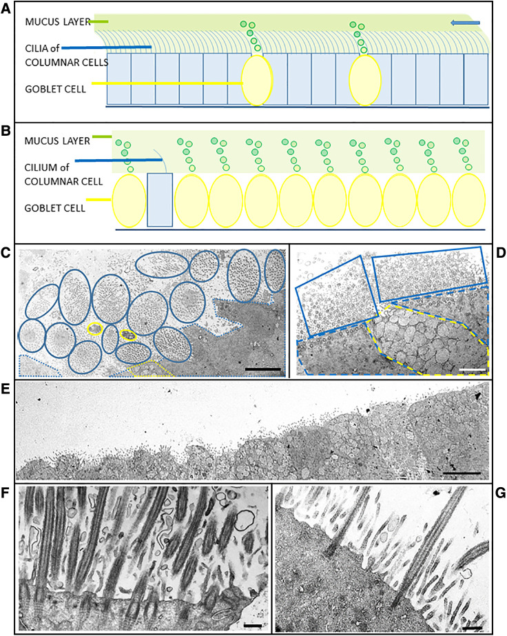
Results: TEM observations confirmed 43% (6) of the clinically-suspected cases (hallmark ultrastructural defects in the dynein arms of the outer doublets), whilst 57% (8) required another PCD testing modality to support ultrastructural observations. Of these, 25% (2) had neither ultrastructural defects nor did they present with bronchiectasis. Of the remaining cases, 83% (5) had very few ciliated cells (all of which were sparsely ciliated), together with goblet cell hyperplasia. There was the apparent absence of ciliary rootlets in 17% (1) case.

Discussion: In resource-limited settings in which TEM is the only available testing modality, confirmatory and probable diagnoses of PCD can be made to facilitate early initiation of treatment of children with chronic respiratory symptoms.

Keywords: South Africa; clinical phenotype; primary ciliary dyskinesia; resource-limited; ultrastructure (electron microscopy).


Conflict of interest statement

The authors declare that the research was conducted in the absence of any commercial or financial relationships that could be construed as a potential conflict of interest.

Figures

Figure 1
Figure 1
Ciliary cross sections (schematic and micrographs) through the median portion of ciliary axonemes from nasal brushings. (A) Normal ultrastructure highlighting noteworthy features used in this study. Each of the nine outer doublets has an outer (green arrow) and an inner (magenta arrow) dynein arm. The doublets are connected to the central pair of microtubules (yellow arrows) by radial spokes (blue arrows). (B) Class 1 defects involving the truncation/absence of outer (green arrows)/inner (magenta arrows) dynein arms. Note a peripheral supernumerary microtubule in the first photomicrograph (black arrow). Not all defects are arrowed. (C) Class 1 defect of microtubular disorganisation (8 peripheral doublets with the ninth doublet in the central area of the axoneme together with the central pair) and missing inner dynein arms (magenta arrows). (D) Secondary ciliary defects involving the central pair of microtubules (either one missing, or an extra one, or none at all distally and in median parts of the axoneme). All cilia are 0.2–0.3 µm in diameter.
Figure 2
Figure 2
Schematics and case micrographs of sections through epithelial cells from nasal brushings. (A) Normal pseudostratified epithelial layer with a 5:1 ratio of multi-ciliated columnar cells to mucin-producing goblet cells. The cilia beat synchronously to move the overlying mucus (and any embedded allergens/pathogens) out of the airways (arrow). (B) Epithelial layer illustrating goblet cell hyperplasia, with rare ciliated columnar cells having reduced numbers of cilia. Lack of ciliation results in accumulation and stasis of the overlying mucus layer. (C) Tangential transverse section through the apices of conical, ciliated columnar cells. Each blue encircled clump of cilia represents one columnar cell. Goblet cells are demarcated in yellow. Although epithelial layers should be planar, the ratio of ciliated:goblet cells is normal. (D) Another tangential section through a goblet cell (yellow dotted line) between two ciliated cells (blue dotted lines). The associated cilia (solid blue lines), which are mostly in cross section, are abundant and evenly spaced. (E) Micrograph illustrating goblet cell hyperplasia (possible case of reduced generation of multiple motile cells) in an epithelial strip in which no ciliated cells are evident. (F) Section through a typical ciliated columnar cell, with numerous ciliary profiles interspersed with microvilli. Note the striated rootlets extending from the basal bodies into the cytoplasm. (G) Section through a columnar epithelial cell illustrating oligocilia - sparsely ciliated with a distinct layer of microvilli. Scale bars: C = 5 µm; D = 2 µm; E = 5 µm; F = 0.5 µm; G = 0.4 µm.

Similar articles

Cited by

References

    1. Machogu E, Gaston B. Respiratory distress in the newborn with primary ciliary dyskinesia. Children. (2021) 8:153. 10.3390/children8020153 - DOI - PMC - PubMed
    1. Legendre M, Zaragosi L-E, Mitchison HM. Motile cilia and airway disease. Semin Cell Dev Biol. (2021) 110:19–33. 10.1016/j.semcdb.2020.11.007 - DOI - PubMed
    1. O’Connor MG, Mosquera R, Metjian H, Marmor M, Olivier KN, Shapiro AJ. Primary ciliary dyskinesia. Chest Pulmonary. (2023) 1:100004. 10.1016/j.chpulm.2023.100004 - DOI
    1. Bhatt R, Hogg C. Primary ciliary dyskinesia: a major player in a bigger game. Breathe. (2020) 16:200047. 10.1183/20734735.0047-2020 - DOI - PMC - PubMed
    1. Goutaki M, Shoemark A. Diagnosis of primary ciliary dyskinesia. Clin Chest Med. (2022) 43(1):127–40. 10.1016/j.ccm.2021.11.008 - DOI - PubMed

LinkOut - more resources