Review
. 2023 Aug 14:16:1231079.
doi: 10.3389/fnmol.2023.1231079. eCollection 2023.

Intrinsic determinants of prion protein neurotoxicity in Drosophila: from sequence to (dys)function

Affiliations
Review

Intrinsic determinants of prion protein neurotoxicity in Drosophila: from sequence to (dys)function

Alessandro Cembran et al. Front Mol Neurosci. .

Abstract

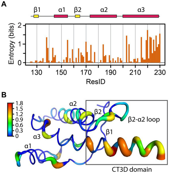
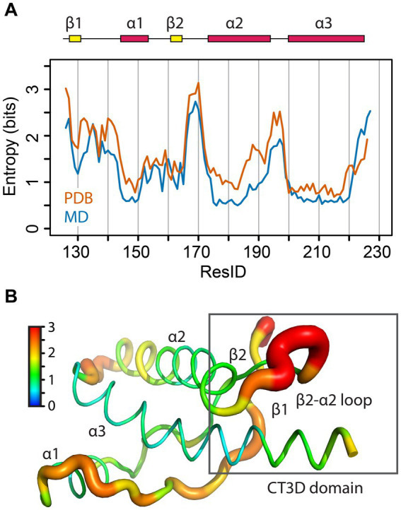
Prion diseases are fatal brain disorders characterized by deposition of insoluble isoforms of the prion protein (PrP). The normal and pathogenic structures of PrP are relatively well known after decades of studies. Yet our current understanding of the intrinsic determinants regulating PrP misfolding are largely missing. A 3D subdomain of PrP comprising the β2-α2 loop and helix 3 contains high sequence and structural variability among animals and has been proposed as a key domain regulating PrP misfolding. We combined in vivo work in Drosophila with molecular dynamics (MD) simulations, which provide additional insight to assess the impact of candidate substitutions in PrP from conformational dynamics. MD simulations revealed that in human PrP WT the β2-α2 loop explores multiple β-turn conformations, whereas the Y225A (rabbit PrP-like) substitution strongly favors a 310-turn conformation, a short right-handed helix. This shift in conformational diversity correlates with lower neurotoxicity in flies. We have identified additional conformational features and candidate amino acids regulating the high toxicity of human PrP and propose a new strategy for testing candidate modifiers first in MD simulations followed by functional experiments in flies. In this review we expand on these new results to provide additional insight into the structural and functional biology of PrP through the prism of the conformational dynamics of a 3D domain in the C-terminus. We propose that the conformational dynamics of this domain is a sensitive measure of the propensity of PrP to misfold and cause toxicity. This provides renewed opportunities to identify the intrinsic determinants of PrP misfolding through the contribution of key amino acids to different conformational states by MD simulations followed by experimental validation in transgenic flies.

Keywords: Drosophila; molecular dynamics; prion protein; protein structure; structure-function.


Conflict of interest statement

The authors declare that the research was conducted in the absence of any commercial or financial relationships that could be construed as a potential conflict of interest.

Figures

Figure 1
Figure 1
Structure of the globular domain of human PrP. (A) Distribution of key pathogenic mutations and the CT3DD. Only a few are identified in different subdomains. (B) Distribution of outstanding substitutions in animals resistant to prion disease. The large letters correspond to human PrP and the smaller correspond to different animals. Y169 is a highly conserved residue with a key role in the dynamics of the CT3DD. Created with PyMOL.
Figure 2
Figure 2
Sequence entropy in the PrP globular domain. (A) The sequence entropy for the PrP structured domain is plotted as a function of the residue number in units of bits. (B) The sequence entropy is mapped onto the human PrP structure, with thicker ribbons corresponding to higher entropy as shown by the color bar.
Figure 3
Figure 3
Amino acid prevalence in the globular domain. Amino acids are colored by their properties, and sizes correspond to their relative frequency for a given position. Generated with WebLogo.
Figure 4
Figure 4
Structural diversity of the CT3DD in human PrP. Structural information for the β2-α2 loop from MD simulations (Myers et al., 2023) was used to created Gibbs free energy isocontours in the space described by the first three principal components calculated using the 𝜑/𝜓 dihedrals. Darker colors correspond to stable regions (see color bar). The pie-chart shows the relative ratio of 310- and β-turn conformations. (A) Human wild-type simulations. The points indicate the center of each of the clusters describing the six main different conformations that were identified. Representative structures of the five β-turns conformations as well as of the 310-turn conformation are shown. (B) The data from human PrP-Y225A MD simulations are plotted using the same approach as above.
Figure 5
Figure 5
Degeneration of mushroom body neurons in Drosophila. (A,D) Control flies expressing the reporter LacZ in mushroom body neurons visualized with CD8-GFP (UAS-LacZ/UAS-CD8-GFP; OK107-Gal4). These clusters contain about 2,000 neurons [(A) The Kenyon cells, Kc] tightly packed in the posterior brain. Their dendritic fields or calyces (Ca) are located underneath the cell bodies. These neurons project to the anterior brain and project into dorsal or medial projections (D). (B,C) 1-day-old flies expressing human PrP-WT (UAS-human PrP-WT/UAS-CD8-GFP; OK107-Gal4) show expansion of the Kc clusters, averaging 30% increased surface compared to controls, whereas flies expressing Y225A (UAS-human PrP-Y225A/UAS-CD8-GFP; OK107-Gal4) show slightly smaller area. (E,F) 35-day-old flies expressing PrP-WT show progressive loss of axonal projections and this loss is minimized in flies expressing Y225A. Representative loop conformations are shown as insets. See Myers et al. (2023) for additional details.
Figure 6
Figure 6
Structural entropy in the globular domain of PrP. (A) The φ/ψ dihedral entropy for the PrP structured domain is plotted in orange as a function of the residue number in units of bits. In blue, the 𝜑/𝜓 dihedral entropy from MD simulations of human PrP is plotted. (B) The φ/ψ dihedral entropy from the PDB data is mapped onto the human PrP structure with thicker ribbons corresponding to higher entropy as shown by the color bar.
Figure 7
Figure 7
Structural diversity of PrP. The structures of the β2-α2 loop from all PrPs available in PBD are mapped onto the axes obtained from principal component analysis of the φ/ψ dihedrals. The markers describe the spread within each PDB set (usually 20 models) as described by the color bar. (A) All 840 PrP structures in PDB are mapped onto the axes obtained from MD simulations for human PrP as reference (Figure 4A; Myers et al., 2023). (B) The 280 PrP-WT structures available in PDB are shown. (C) The 280 mouse PrP (WT and mutant) structures available in PDB are reported. The structure of the β2-α2 loop for Y169G is shown as a cartoon. (D) The complete ensemble of 20 PDB structures for selected PrPs are shown to illustrate the population of specific conformations (mule deer, horse, mouse Y169G) or their conformational diversity (human).

References

    1. Alonso D. O., DeArmond S. J., Cohen F. E., Daggett V. (2001). Mapping the early steps in the pH-induced conformational conversion of the prion protein. Proc. Natl. Acad. Sci. U. S. A. 98, 2985–2989. doi: 10.1073/pnas.061555898, PMID: - DOI - PMC - PubMed
    1. Apetri A. C., Surewicz K., Surewicz W. K. (2004). The effect of disease-associated mutations on the folding pathway of human prion protein. J. Biol. Chem. 279, 18008–18014. doi: 10.1074/jbc.M313581200, PMID: - DOI - PubMed
    1. Apetri A. C., Vanik D. L., Surewicz W. K. (2005). Polymorphism at residue 129 modulates the conformational conversion of the D178N variant of human prion protein 90-231. Biochemistry 44, 15880–15888. doi: 10.1021/bi051455+, PMID: - DOI - PubMed
    1. Appleby B. S., Shetty S., Elkasaby M. (2022). Genetic aspects of human prion diseases. Front. Neurol. 13:1003056. doi: 10.3389/fneur.2022.1003056, PMID: - DOI - PMC - PubMed
    1. Barlow R. M., Rennie J. C. (1976). The fate of ME7 scrapie infection in rats, guinea-pigs, and rabbits. Res. Vet. Sci. 21, 110–111. - PubMed

LinkOut - more resources