[Preprint]. 2023 Aug 17:2023.08.16.553618.
doi: 10.1101/2023.08.16.553618.

Structure of C. elegans TMC-2 complex suggests roles of lipid-mediated subunit contacts in mechanosensory transduction

Affiliations

Structure of C. elegans TMC-2 complex suggests roles of lipid-mediated subunit contacts in mechanosensory transduction

Sarah Clark et al. bioRxiv. .

Update in

Abstract

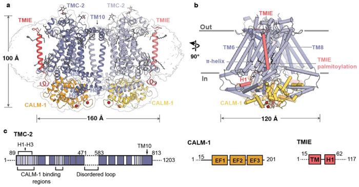
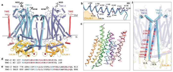
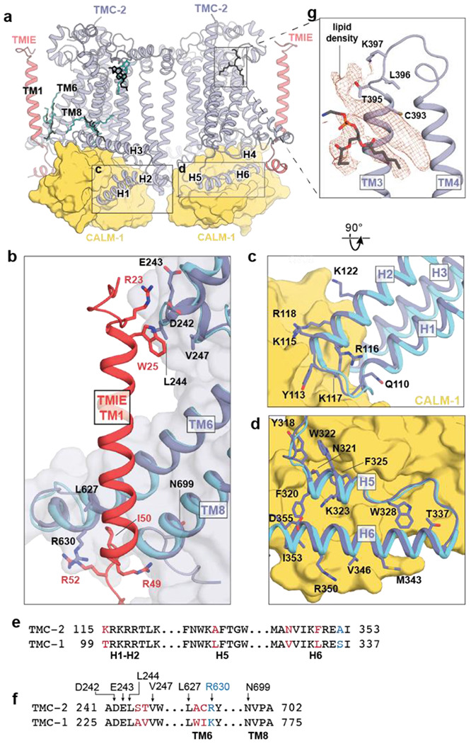
Mechanotransduction is the process by which a mechanical force, such as touch, is converted into an electrical signal. Transmembrane channel-like (TMC) proteins are an evolutionarily-conserved family of ion channels whose function has been linked to a variety of mechanosensory processes, including hearing and balance sensation in vertebrates and locomotion in Drosophila. The molecular features that tune homologous TMC ion channel complexes to diverse mechanical stimuli are unknown. Caenorhabditis elegans express two TMC homologs, TMC-1 and TMC-2, both of which are the likely pore-forming subunits of mechanosensitive ion channels but differ in their expression pattern and functional role in the worm. Here we present the single particle cryo-electron microscopy structure of the native TMC-2 complex isolated from C. elegans. The complex is composed of two copies each of the pore-forming TMC-2 subunit, the calcium and integrin binding protein CALM-1 and the transmembrane inner ear protein TMIE. Comparison of the TMC-2 complex to the recently published cryo-EM structure of the C. elegans TMC-1 complex reveals differences in subunit composition and highlights conserved protein-lipid interactions, as well as other structural features, that together suggest a mechanism for TMC-mediated mechanosensory transduction.


Conflict of interest statement

Competing Interest Statement: The authors declare no competing interests.

Figures

Figure 1:
Figure 1:. Architecture of the TMC-2 complex.
a, Overall architecture of the TMC-2 complex, viewed parallel to the membrane. TMC-2 (dark and light purple), CALM-1 (orange and yellow), and TMIE (dark and light pink), are shown in a cartoon diagram. Lipid-like molecules and N-glycans are colored grey, while calcium ions red. The map refined without a mask is shown as a transparent envelope. b, A 90° rotated-view of the TMC-2 complex, viewed parallel to the membrane. Structural features of interest are indicated. α-Helices are shown as cylinders. c, Schematic representation of proteins that form the TMC-2 complex. For TMC-2, helices are shown in light purple and transmembrane helices are shown in dark purple. Dashed lines indicate regions that were not observed in the cryo-EM structure. H = helix, TM = transmembrane domain, EF1-EF3 = EF-hand domains.
Figure 2:
Figure 2:. The TMC-1 and TMC-2 complexes have similar architectures, yet adopt distinct conformations at the dimer interface.
a, Superposition of backbone atoms of the TMC-2 complex with backbone atoms of one protomer of the TMC-1 complex, shown on the left. TMC-1 is colored cyan and the CALM-1 and TMIE auxiliary subunits from the TMC-1 complex are colored yellow and pink, respectively. The TMC-2 complex is colored as in Figure 1. N-glycan molecules are colored black and calcium ions are colored dark red. b, TMC-2 helix H3, shown in purple, is superposed with TMC-1 helix H3, shown in cyan. Residues that confer an amphipathic nature upon the TMC-2 H3 helix are shown as sticks. c, The TMC-1 transmembrane helices (TM1-TM9), shown in grey, superposed on the TMC-2 transmembrane helices, shown in different colors, using backbone carbon atoms. d, Sequence alignment of the amino acid residues of the TMC-2 and TMC-1 H3 helices. Residues are colored black for identical, blue for conservatively substituted, or red for non-conservatively substituted. e, Sequence alignment of amino acid residues of the TMC-2 and TMC-1 TM10 regions. Residues are colored as in (d). f, Closeup view of the dimer interface of the superimposed complex from panel (a), rotated ~45° about an axis perpendicular to the membrane plane, highlighting the different conformations of the TM10 regions in the two complexes. The positions of the non-conserved residues in TM10 of TMC-2, compared to TMC-1 and as defined in panel (e), are shown as red bands.
Figure 3:
Figure 3:. TMC-1 and TMC-2 share a conserved interaction interface with the auxiliary subunits TMIE and CALM-1.
a, Lipids with strong density features and similar locations in the TMC-1 and TMC-2 density maps are shown in the context of the TMC-2 complex. TMC-1 lipids are cyan and TMC-2 lipids are black. b, The subunit interface of TMIE in the TMC-1 or TMC-2 complex. TMC-2 is purple, TMC-1 is cyan, and TMIE is red. Interacting residues are shown as sticks. c, The subunit interface of CALM-1 with TMC-1 or TMC-2 helices H1-H2. CALM-1 is colored yellow and interacting residues are shown as sticks. d, The subunit interface of CALM-1 with TMC-1 or TMC-2 helices H5-H6, colored as in (c). e, Sequence alignment of the residues from the cytoplasmic helices of TMC-1 and TMC-2 that form the interaction interface with CALM-1. Residues in black are conserved. Residues in blue are conservatively substituted and residues in red are not conserved. f, Sequence alignment of TMC-2 and TMC-1 residues that interact with TMIE. Residues are colored as in (e). g, Lipid-like density features observed in the TMC-2 map that were not observed in the TMC-1 map. Density map is shown in red and TMC-2 is shown in purple.
Figure 4:
Figure 4:. The putative ion conduction pore of TMC-2.
a, The putative location of the pore is shown as gold mesh. The pore-lining helices, TM4-8, are shown in purple and the remaining TM helices are shown in white. b, An expanded view of the possible ion permeation pathway, showing pore-lining residues as sticks. Residues that are not conserved between TMC-1 and TMC-2 are shown in red. A π-helical turn within TM6 of TMC-2 is shown in orange. c, Radii of the TMC-1 and TMC-2 pores plotted along the pore axis, calculated by Mole 2.5. TMC-1 is shown in blue and TMC-2 is shown in purple. d, Hydropathy plot of the TMC-1 pore (left) and TMC-2 pore (right) calculated by Mole 2.5. Regions of the pore with high hydrophobicity values are colored yellow and regions with high hydrophilicity values are colored blue. The size of the pore is plotted along the pore axis. e, Multiple sequence alignment of selected residues from the putative TMC-2 and TMC-1 pore-lining helices of different species, with the residues colored as in panel (b).
Figure 5:
Figure 5:. Lipid-mediated interactions between TMIE and TMC-1/2.
a, Palmitoylation of TMIE C43 and C44 is observed in the TMC-2 complex. Interacting residues are shown as sticks and a phospholipid is shown in light grey. Cryo-EM density is shown as red mesh, at a contour level of 5.68 RMSD. b, Palmitoylation of TMIE C43 and C44 is also observed in the TMC-1 complex. TMC-1 is shown in cyan and other coloring is depicted as in (a). The density map contour level is 4.62 RMSD. c, Schematic illustrating how lipid-mediated interactions between TMIE and the TMC-1/2 transmembrane helices may translate forces to ion channel gating. Lipid interactions may stabilize the closed conformation of the channel (left). Disruption of the membrane due to mechanical forces could alter these lipid interactions, leading to ion channel opening (right). Phospholipid head groups are colored yellow.

References

    1. Kefauver J. M., Ward A. B., Patapoutian A., Discoveries in structure and physiology of mechanically activated ion channels. Nature 587, 567–576 (2020). - PMC - PubMed
    1. Pan B. et al., TMC1 and TMC2 are components of the mechanotransduction channel in hair cells of the mammalian inner ear. Neuron 79, 504–515 (2013). - PMC - PubMed
    1. Kurima K. et al., TMC1 and TMC2 Localize at the Site of Mechanotransduction in Mammalian Inner Ear Hair Cell Stereocilia. Cell reports 12, 1606–1617 (2015). - PMC - PubMed
    1. Chou S. W. et al., A molecular basis for water motion detection by the mechanosensory lateral line of zebrafish. Nature communications 8, 2234 (2017). - PMC - PubMed
    1. Pacentine I. V., Nicolson T., Subunits of the mechano-electrical transduction channel, Tmc1/2b, require Tmie to localize in zebrafish sensory hair cells. PLoS Genet 15, e1007635 (2019). - PMC - PubMed

Publication types