[Preprint]. 2023 Aug 17:2023.08.16.553624.
doi: 10.1101/2023.08.16.553624.

Triaging of α-helical proteins to the mitochondrial outer membrane by distinct chaperone machinery based on substrate topology

Affiliations

Triaging of α-helical proteins to the mitochondrial outer membrane by distinct chaperone machinery based on substrate topology

Gayathri Muthukumar et al. bioRxiv. .

Update in

Abstract

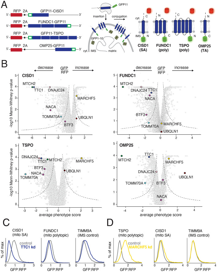
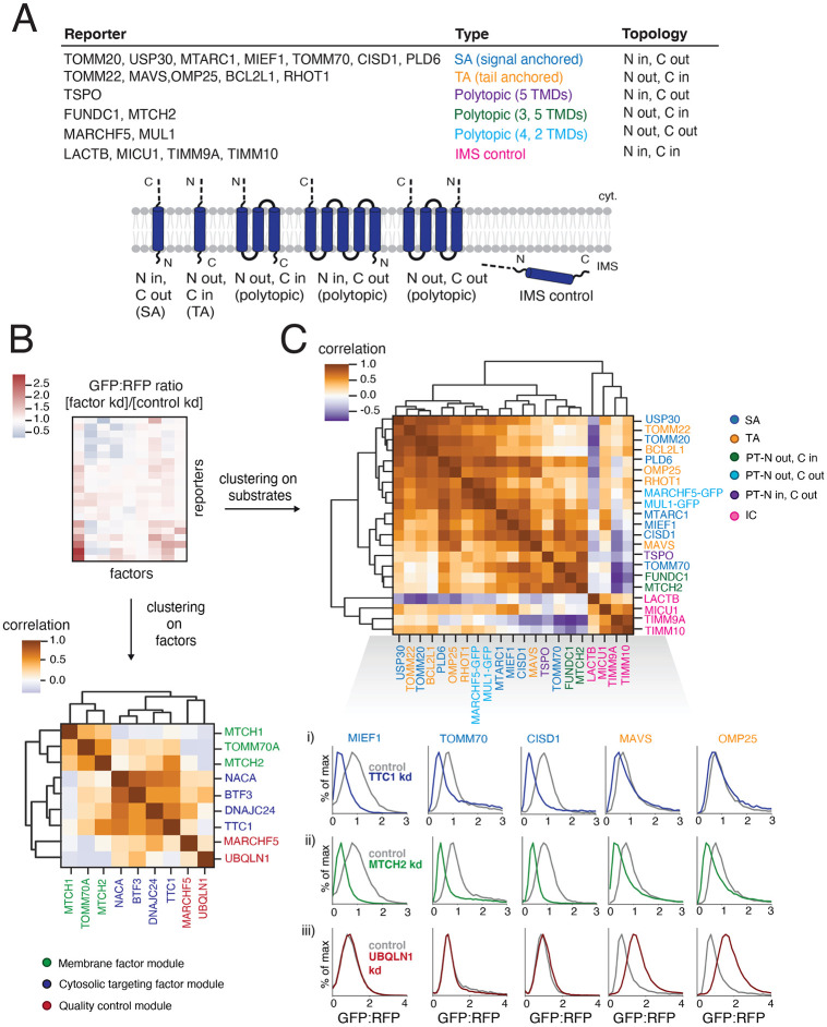
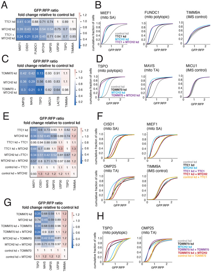
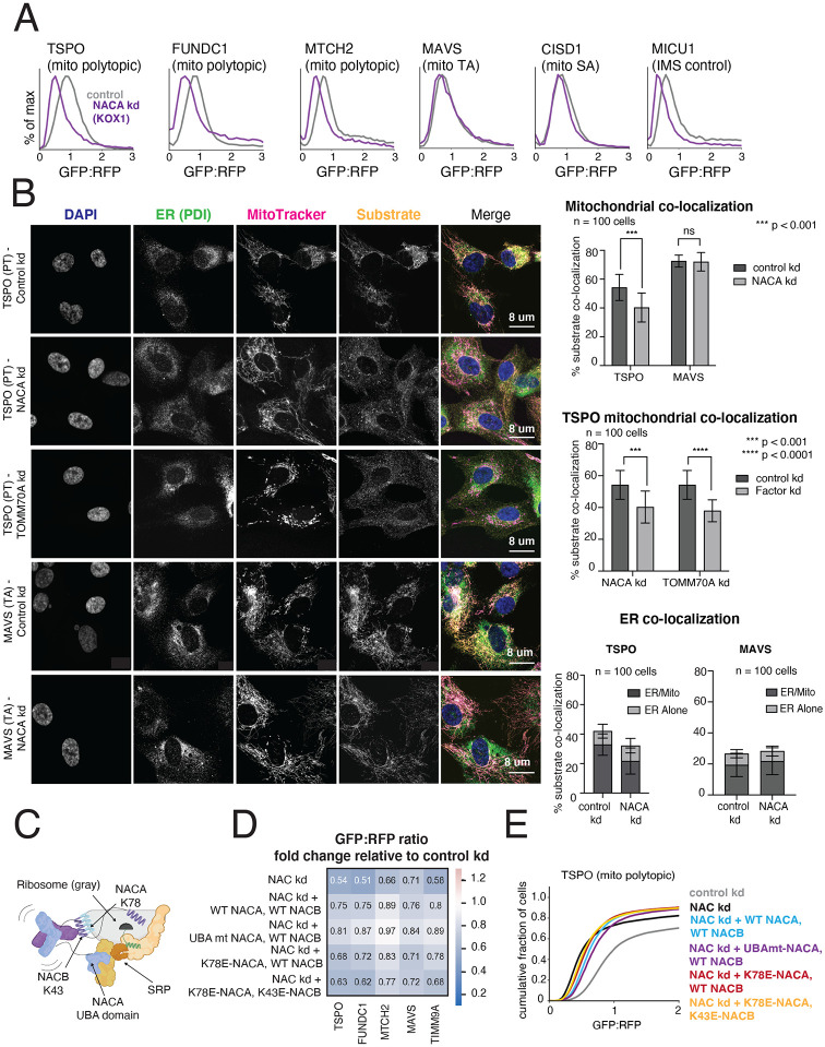
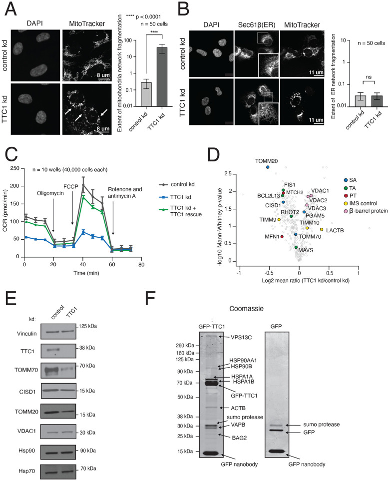
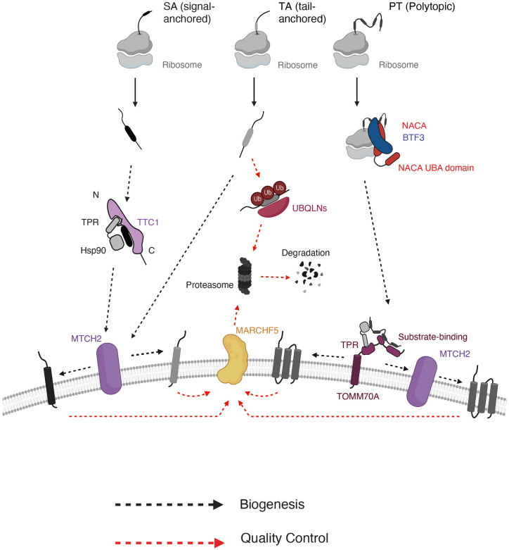
Mitochondrial outer membrane α-helical proteins play critical roles in mitochondrial-cytoplasmic communication, but the rules governing the targeting and insertion of these biophysically diverse substrates remain unknown. Here, we first defined the complement of required mammalian biogenesis machinery through genome-wide CRISPRi screens using topologically distinct membrane proteins. Systematic analysis of nine identified factors across 21 diverse α-helical substrates reveals that these components are organized into distinct targeting pathways which act on substrates based on their topology. NAC is required for efficient targeting of polytopic proteins whereas signal-anchored proteins require TTC1, a novel cytosolic chaperone which physically engages substrates. Biochemical and mutational studies reveal that TTC1 employs a conserved TPR domain and a hydrophobic groove in its C-terminal domain to support substrate solubilization and insertion into mitochondria. Thus, targeting of diverse mitochondrial membrane proteins is achieved through topological triaging in the cytosol using principles with similarities to ER membrane protein biogenesis systems.

Keywords: CRISPR; NAC; TTC1; biogenesis pathway organization; cell biology; chaperone complexes; cytosolic targeting; genetic screens; topology; α-helical outer mitochondrial membrane proteins.


Conflict of interest statement

Declaration of interests: J.S.W. declares outside interest in 5 AM Venture, Amgen, Chroma Medicine, KSQ Therapeutics, Maze Therapeutics, Tenaya Therapeutics, Tessera Therapeutics, and Third Rock Ventures. R.M.V. is a consultant and equity holder in Gate Bioscience.

Figures

Figure 1.
Figure 1.. Identifying factors required for the biogenesis of topologically distinct α-helical mitochondrial outer membrane proteins.
(A) The insertion of α-helical mitochondrial outer membrane proteins of diverse topologies (SA: signal-anchored, poly: polytopic and TA: tail-anchored) was queried using a split GFP reporter system. GFP11-fused reporters were expressed in a K562 cell line constitutively expressing GFP1-10 in the intermembrane space (IMS) such that insertion in the correct topology would result in complementation and GFP fluorescence. RFP (red fluorescent protein) separated by a viral P2A acts as a translation normalization marker. (B) Volcano plots of the GFP:RFP stabilization phenotype for the three strongest sgRNAs and Mann-Whitney p-values for the genome-wide CRISPRi FACS screens (two independent replicates each) for the four reporters in (A). Each gene is indicated by a gray dot, with specific genes of interest that either decrease or increase the GFP:RFP ratio highlighted across all four screens. The OMP25 screen was conducted in a previous study with identical screening conditions and is replotted to allow for direct comparison (Guna et. al, 2022). (C) Integration of the indicated GFP11-fused reporters in K562 cell lines expressing IMS GFP1-10 in the presence of a non-targeting (control) sgRNA or sgRNAs targeting TTC1 or (D) MARCHF5. TIMM9A is an IMS control reporter. Flow data was normalized based on the GFP:RFP ratio and plotted as histograms. See Figures S1 and S2.
Figure 2.
Figure 2.. Systematically exploring pathways affecting the biogenesis of diverse α-helical mitochondrial outer membrane proteins
(A) Table describing a comprehensive set of α-helical outer membrane proteins used as reporters to query the specificity of hits (highlighted in Figure 1B) derived from the genome-wide CRISPRi screens. Type and topology of each protein is stated and depicted. IMS control reporters were also included (for a total of 21 substrates assayed). ‘LACTB’ and ‘MICU1’ indicate just the respective targeting sequence fused to GFP11. For proteins with both termini in the cytosol (MUL1 and MARCHF5) and therefore incompatible with the split GFP system, full-length GFP was fused to the C-termini. (B) To analyze the effects of factors of interest on α-helical proteins of different topologies, each reporter in (A) was expressed in K562 ZIM3 CRISPRi cells constitutively expressing IMS GFP1-10 and a sgRNA guide against the factor or a non-targeting control and analyzed by flow cytometry. The GFP:RFP ratios were then normalized to the non-targeting control ratios to generate a comprehensive dataset represented as a heatmap (see Figure S3A for details). Hierarchical clustering of the correlation matrix of all factors results in the assignment of putative biogenesis pathways. (C) Clustering of the correlation matrix of all reporters assigns outer membrane reporters into clusters largely defined by type and topology. Individual histograms showing GFP:RFP changes in response to factor depletion that are predictive of SA and TA reporter clustering patterns are shown here: i) responses to TTC1 depletion, ii) responses to MTCH2 depletion, iii) responses to UBQLN1 depletion. See Figure S3 for complete reporter GFP:RFP changes (for all factors). See Figures S3 and S4.
Figure 3.
Figure 3.. TTC1 and TOMM70 exert effects on α-helical membrane protein integration independent of their effects on the MTCH2 insertase
(A) Arrayed dual guides were used to assess the genetic interaction of TTC1 and MTCH2 by monitoring outer membrane integration of GFP11-fused α-helical reporters dependent on TTC1 and MTCH2 (MIEF1, CISD1, MTCH2), MTCH2 alone (USP30, FUNDC1, OMP25) and neither (TSPO, TIMM9A) (dependencies noted from Figure S3A). Reporters were expressed in K562 ZIM3 CRISPRi IMS GFP1-10 cells and i) sgRNAs targeting TTC1 and MTCH2, ii) sgRNAs targeting TTC1 and a non-targeting control, iii) sgRNAs targeting MTCH2 and a non-targeting control. Data is represented as a heatmap colored by the fold change in reporter integration (GFP:RFP ratio) for each condition relative to cells expressing a non-targeting sgRNA. (B) Flow data for representative reporters from (A) plotted as cdf (cumulative distribution function) plots. (C) The genetic interaction of TOMM70A and MTCH2 was assessed in a similar manner to (A) with reporters dependent on TOMM70A and MTCH2 (OMP25, MAVS), TOMM70A alone (TSPO) and neither (MICU1, LACTB, TIMM9A). As in (A), dependencies are noted from arrayed screen results in Figure S3A. (D) Flow data for representative reporters from (C) plotted as cdf plots. (E) To determine the extent to which TTC1 biogenesis defects are a secondary consequence of MTCH2 loss, outer membrane integration of GFP11-fused α-helical reporters was measured in K562 IMS GFP1-10 cells constitutively expressing a sgRNA against TTC1 and i) exogenous 3xFLAG-tagged TTC1 in a cassette with a BFP translation marker, ii) exogenous MTCH2 with a BFP translation marker and iii) BFP alone. Reporter integration was measured with the same conditions for cells expressing sgRNAs targeting MTCH2 and non-targeting sgRNAs (control). Data is represented as a heatmap colored in the same manner as (A) and (C). (F) Flow data for representative reporters from (E) plotted as cdf plots. (G) The extent to which TOMM70A biogenesis defects are a secondary consequence of MTCH2 loss is assessed in the same manner as (E). (H) Flow data for representative reporters from (E) plotted as cdf plots. See Figure S5.
Figure 4.
Figure 4.. The NAC complex is required for the efficient delivery of polytopic α-helical proteins to the mitochondrial outer membrane
(A) Integration of indicated GFP11-fused SA, TA, polytopic and IMS control reporters in K562 KOX1 CRISPRi cells constitutively expressing IMS GFP1-10 and either a sgRNA targeting NACα or a non-targeting sgRNA (control). (B) (Left) Changes in localization of endogenous TSPO (polytopic) and MAVS (TA) were assessed using immunostaining and confocal microscopy in RPE1 ZIM3 CRISPRi cells expressing sgRNAs targeting NACα, TOMM70 or a non-targeting control. Merge colors: Blue: DAPI, Green: ER, Yellow: Substrate, Magenta: MitoTracker. (Right) Quantification of endogenous substrates co-localized to MitoTracker (mitochondrial marker) and PDI (ER marker) were measured and plotted (see Methods for details). Quantification of substrates co-localized to the ER were split into i) percent co-localized to ER/MitoTracker overlapping regions and ii) percent co-localized to the rest of the ER (‘ER alone’) (see FIgure S6D). For more detailed substrate localization analysis across all conditions, see Figures S6C-F. Error bars show mean ± SD of 100 cells. Statistical significance was evaluated by multiple unpaired t-tests with the Holm-Sidak multiple test correction. ***, p < 0.001. ****, p < 0.0001. ns (non-significant), p > 0.05. (C) Model of a translating ribosome engaging NAC and SRP. NACα (blue) and NACβ (purple) interact with the ribosome (gray) through electrostatic interactions mediated by the indicated lysine residues (NACα K78 and NACβ K43). SRP is recruited to the ribosome through electrostatic interactions with the UBA domain of NACα (at residues D205 and N208), where it engages the substrate nascent chain (green) co-translationally. (D) The effects of ribosome-binding mutants of NACα and NACβ (K78E and K43E respectively) and a UBA-domain binding mutant of NACα (D205R + N208R) on integration of select GFP11-fused reporters in K562 KOX1 CRISPRi IMS GFP1-10 cells was tested using a similar strategy as in Figures 3E and 3G. Both members of the NAC complex were simultaneously depleted using CRISPRi and abilities of exogenous wild-type (WT) and mutant versions of NACα and NACβ to rescue reporter integration defects were assessed. Rescue constructs are in a cassette with a BFP translation marker. Data is represented as a heatmap colored by the effects of exogenous additions of NACα and NACβ (WT and mutant versions as indicated) on reporter integration in the mitochondria relative to the control condition (non-targeting sgRNA + BFP alone, as shown in Figures 3E and 3G). (E) Flow cytometry data for polytopic reporter TSPO across all conditions shown in (D) represented as a cdf plot. See Figures S6 and S7.
Figure 5.
Figure 5.. TTC1 is required for mitochondrial integrity and the biogenesis of endogenous outer membrane proteins
(A) RPE1 ZIM3 CRISPRi cells expressing either a sgRNA targeting TTC1 or a non-targeting sgRNA were analyzed by super-resolution confocal microscopy to assess changes in mitochondrial morphology. The white arrows indicate mitochondrial fragmentation in TTC1 depleted cells. Extent of network fragmentation is calculated using the median count: area ratio of the mitochondrial network in each condition with log10 values plotted. (B) The effects of TTC1 depletion on ER morphology in RPE1 ZIM3 CRISPRi cells, assessed through immunostaining with Sec61β and calculated as in (A). For (A) and (B), error bars show mean ± SD of 50 cells. Statistical significance was evaluated using the Welch corrected unpaired t-test. ****, p < 0.0001. ns (non-significant), p > 0.05. (C) Mitochondrial respiration was assessed in K562 ZIM3 CRISPRi cells depleted of TTC1 where levels were then rescued with an exogenous copy (see Figure S10D for protein levels) using a Seahorse assay to monitor oxygen consumption rates (OCR). Arrows indicate time of drug treatment. Data are presented as average ± SD, n = 10. (D) TMT mass-spectrometry analysis of K562 KOX1 CRISPRi cells expressing guides targeting TTC1 relative to cells expressing non-targeting guides (control). Data shown here are normalized to mitochondrial proteome levels and evaluated for statistical significance across 3 biological replicates. Outer membrane proteins of interest are colored by topology. (E) Immunoblotting of endogenous proteins in K562 KOX1 CRISPRi TTC1 depleted cells and control cells (non-targeting sgRNA) using vinculin as the loading control. (F) HEK293T cells stably expressing endogenously tagged TTC1-GFP were used to identify endogenous TTC1 interaction partners. TTC1-GFP was purified under native conditions using an anti-GFP nanobody (Pleiner et al, 2020). Interaction partners were analyzed by separation through SDS-PAGE, detection with Coomassie staining and identification by mass spectrometry. As a control for non-specific interactions, HEK293T cells were transduced with GFP before purification using the GFP-nanobody and downstream analysis in the same manner. See Figure S8.
Figure 6.
Figure 6.. TTC1 chaperones nascent SA proteins and promotes insertion into mitochondria in vitro, using a C-terminal hydrophobic groove to direct biogenesis
(A) Indicated substrates tagged with 3xFLAG were translated in rabbit reticulocyte lysate (RRL) in the presence of 35S and released with puromycin. Immunoprecipitation and immunoblotting show the levels of TTC1 associated with the nascent substrates. (B) AlphaFold2 model of TTC1 structure colored by i) evolutionary conservation using the ConSurf server (see Methods for details) showing high conservation in the TPR domain and the C-terminus (left) and ii) electrostatic potential indicating a hydrophobic groove at the C-terminus (right). (C) The effects of putative heat-shock binding TPR domain mutants and C-terminal point mutants (individual residues indicated in (B)) on integration of indicated GFP11-fused reporters in K562 ZIM3 CRISPRi IMS GFP1-10 cells expressing a sgRNA targeting TTC1 were assessed in a similar manner to Figure 4D. (Left) Data is represented as a heatmap colored by the fold change in reporter integration (GFP:RFP ratio) for TTC1 depleted cells expressing WT or mutant TTC1. (Right) Flow data for the effects of the R190A+R194A TPR domain and I271D C-terminal mutants on SA reporter CISD1 is plotted as a cdf plot. (D) CISD1 was translated in vitro in the presence of 35S in the PURE system in the presence of either i) no chaperone, ii) purified calmodulin, iii) purified WT TTC1, iv) purified I271D-TTC1. The translations were run on a sucrose gradient and the extent of the substrate remaining soluble was determined by autoradiography. (D) (Left) Insertion into mitochondria isolated from wild-type K562 cells of endogenous CISD1 (SA), OMP25 (TA) and Su9-DHFR translated from RRL in the presence of increasing amounts of WT TTC1 or the I271D mutant (matched concentrations). Insertion is assessed by PK (proteinase K) digestion followed by an IP of the protected 6xHis tag. (Right) Quantification of the fold increase for mitochondrial insertion of OMP25 and CISD1 stimulated by WT and I271D-TTC1. See Figures S9 and S10.
Figure 7.
Figure 7.. Working model for α-helical outer membrane protein biogenesis using multiple parallel pathways.
Biogenesis pathways including cytosolic targeting, membrane insertion and quality control are depicted for three main types of α-helical outer membrane proteins (signal-anchored, tail-anchored and polytopic). Nascent outer membrane proteins are likely triaged during targeting in the cytosol, with signal-anchored proteins being targeted through cytosolic chaperone TTC1 and polytopic proteins targeted through co-translational chaperone complex NAC. Whether NAC directly engages and targets the polytopic proteins to the OMM remains to be explored. All proteins converge on MTCH1/2 insertases at the membrane for insertion, with some polytopic proteins depending more heavily on recruitment by TOMM70. Similarly, membrane-resident E3 ligase MARCHF5 handles quality control of aberrant/misfolded TMDs for substrates of all α-helical protein types, with some variation within protein type. In the cytosol, quality control processes are more divergent with tail-anchored proteins degraded using UBQLNs and no definitive pathway for other protein types.

References

    1. von Heijne G. (2007). The membrane protein universe: what’s out there and why bother?) J. Intern. Med. 261, 543–557. - PubMed
    1. Rapoport T.A., Li L., Park E. (2017). Structural and Mechanistic Insights into Protein Translocation. Annu. Rev. Cell. Dev. Biol. 33, 369–390. - PubMed
    1. Hegde R.S. and Keenan R.J. (2022). The mechanisms of integral membrane protein biogenesis. Nat. Rev. Mol. Cell. Biol. 23, 107–124. - PubMed
    1. Krogh A., Larsson B., von Heijne G. and Sonnhammer E. L. (2001). Predicting transmembrane protein topology with a hidden Markov model: application to complete genomes. J. Mol. Biol. 305, 567–580. - PubMed
    1. Wickner W. and Schekman R. (2005). Protein translocation across biological membranes. Science 310, 1452–1456. - PubMed

Publication types