[Preprint]. 2023 Aug 17:2023.08.17.553660.
doi: 10.1101/2023.08.17.553660.

Structural elucidation of recombinant Trichomonas vaginalis 20S proteasome bound to covalent inhibitors

Affiliations

Structural elucidation of recombinant Trichomonas vaginalis 20S proteasome bound to covalent inhibitors

Jan Silhan et al. bioRxiv. .

Update in

Abstract

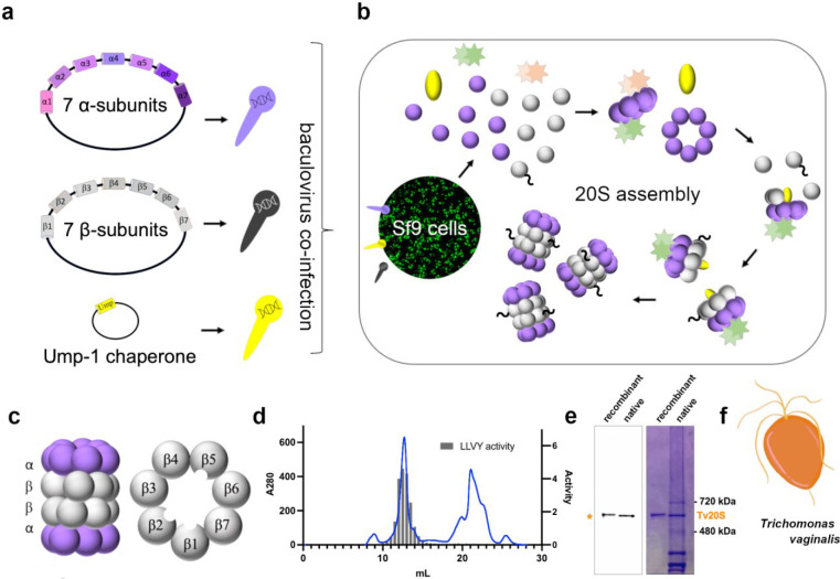
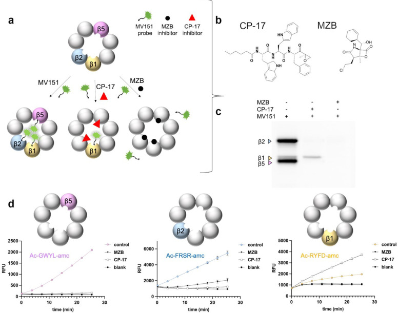
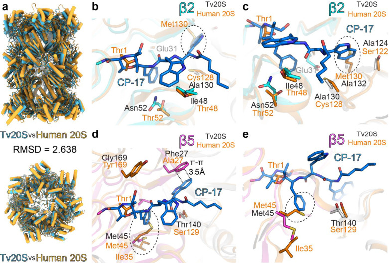
Proteasomes are essential for protein homeostasis in mammalian cells1-4 and in protozoan parasites such as Trichomonas vaginalis (Tv).5 Tv and other protozoan 20S proteasomes have been validated as druggable targets.6-8 However, in the case of Tv 20S proteasome (Tv20S), biochemical and structural studies were impeded by low yields and purity of the native proteasome. We successfully made recombinant Tv20S by expressing all seven α and seven β subunits together with the Ump-1 chaperone in insect cells. We isolated recombinant proteasome and showed that it was biochemically indistinguishable from the native enzyme. We confirmed that the recombinant Tv20S is inhibited by the natural product marizomib (MZB)9 and the recently developed peptide inhibitor carmaphycin-17 (CP-17)8,10. Specifically, MZB binds to the β1, β2 and β5 subunits, while CP-17 binds the β2 and β5 subunits. Next, we obtained cryo-EM structures of Tv20S in complex with these covalent inhibitors at 2.8Å resolution. The structures revealed the overall fold of the Tv20S and the binding mode of MZB and CP-17. Our work explains the low specificity of MZB and higher specificity of CP-17 towards Tv20S as compared to human proteasome and provides the platform for the development of Tv20S inhibitors for treatment of trichomoniasis.


Conflict of interest statement

Conflict of Interests The authors declare no conflict of interests.

Figures

Fig. 1:
Fig. 1:. Cloning, Recombinant Expression, and Purification of Tv20S Proteasome.
a) Cloning of three vectors containing either 7 α-subunits, 7 β-subunits or Ump-1 into baculovirus. b) Co-infection of SF9 cells with baculovirus for proteasome expression and assembly. c) Side view structure of the 20S proteasome complex showing two β rings sandwiched between two α rings. Planer view of the β ring showing the three catalytic subunits located within the central tunnel of the proteasome. d) Final purification step using Superose 6 chromatography. Absorbance at 280 nm (blue line) and proteasome activity assessed using a proteasome-specific fluorogenic substrate Suc-LLVY-amc (grey bar chart). MV151 and Coomassie blue-stained native PAGE showing the purity of the recombinantly purified proteasome complex compared to native Tv20S isolated from T. vaginalis. F) Cartoon representation of T. vaginalis at 100x magnification.
Fig. 2:
Fig. 2:. Characterization of the proteasome-inhibitor interaction and inhibition kinetics.
a) The figure illustrates the schematic representation of active sites during inhibition by a MZB or CP-17, followed by visualization through fluorescent MV151 probing. The inhibitor is depicted binding to specific active sites, thereby blocking enzymatic activity. Subsequent visualization techniques provide insights into the binding interaction and potential conformational changes induced by the inhibitor. b) Chemical structure of MZB and CP-17. c) rTv20S was first incubated with DMSO, 50 μM CP-17 or 50 μM inhibitor for 1 hour, and then incubated with 2 μM MV151 for 16 hours and the catalytic subunits were imaged on a denaturing gel at 470 nm excitation 530 nm emission. d) Screening and validation of inhibitor specificity using subunit-specific proteasome substrates.
Fig. 3:
Fig. 3:. Cryo-EM structures inhibitors MZB and CP-17 bound active sites of Tv20S proteasome.
The atomic models of Tv20S with covalent inhibitors a MZB or e CP-17. The panels a and e, the cryo-electron map highlights the inhibitors in active sites (shown as green mesh), while catalytic units are depicted in different colors (β1 yellow, β2 cyan and β5 magenta). The electron map for MZB was observed only in two catalytic sites β1 and β2 (panels b&c), on the other hand the electron map for CP-17 was observed only in catalytic sites β2 and β5 (panels f&g). These four panels (b,c,f,g) show a stick representation of inhibitors (orange and blue) covalently linked to active sites threonine residues. Amino acid residues within a reach of non-covalent interaction (up to 4 Å) are shown as sticks. Panels d and h show depict the surface representation and structural orchestration of the active site pockets and their connection within Tv20S proteasome with the inhibitors bound. The dashed line outlines the C2 symmetry.
Fig. 4:
Fig. 4:. Comparison of Tv20S and human 20S structures:
The panel a comparison of human (orange) and Trichomonas vaginalis (light blue) 20S proteasomes. The further panels show structural overlay of human 20S and covalent inhibitor CP-17 (blue sticks) in the active site pockets of β2 (panels b&c) and β5 (panels d&e) of Tv20S. The residues responsible for major structural differences in these active sites are shown as sticks (human-orange, Tv20S β2 cyan and β5 magenta, other subunits shown in white). The predicted clashes between CP-17 and amino acids from human 20S are encircled with dotted line. In panels b&c, Met130 of human β2 is in clash with one of the indole rings of CP-17, whilst in Tv20S this moiety is accommodated in a deep hydrophobic pocket formed by Ala132 and Ala130. Similarly, in panels d&e, the pocked formed by Met45 of Tv20S β5 allows sufficient space for phenyl ring of CP-17. In this location human β5 Met45 is pushed by Ile35 towards the active site, forming a potential barrier for CP-17.
Fig. 5:
Fig. 5:. Surface detail of comparison of Tv20S and human 20S active sites.
Tv20S surface is in light blue. Human 20S proteasomes is in light yellow and its Thr1 is shown as orange sticks. MZB and CP-17 are shown as blue sticks is displayed in human active site pockets using structural overlay from Fig.4. The clashes are encircled with a red dotted line. In panel h, Met130 of human β2 forms shallower binding pocket in this region whist similar elevation of active site pocket is observed formed by Met45 from human β5. In this location human β5 Met45 in this ligand-free structure is pushed by Ile35 towards the active site.

Similar articles

References

    1. Finley D. Recognition and processing of ubiquitin-protein conjugates by the proteasome. Annual review of biochemistry 78, 477–513 (2009). 10.1146/annurev.biochem.78.081507.101607 - DOI - PMC - PubMed
    1. Collins G. A. & Goldberg A. L. The Logic of the 26S Proteasome. Cell 169, 792–806 (2017). 10.1016/j.cell.2017.04.023 - DOI - PMC - PubMed
    1. Lecker S. H., Goldberg A. L. & Mitch W. E. Protein degradation by the ubiquitin-proteasome pathway in normal and disease states. J Am Soc Nephrol 17, 1807–1819 (2006). 10.1681/ASN.2006010083 - DOI - PubMed
    1. Majumder P. & Baumeister W. Proteasomes: unfoldase-assisted protein degradation machines. Biological chemistry 401, 183–199 (2019). 10.1515/hsz-2019-0344 - DOI - PubMed
    1. Munoz C., San Francisco J., Gutierrez B. & Gonzalez J. Role of the Ubiquitin-Proteasome Systems in the Biology and Virulence of Protozoan Parasites. Biomed Res Int 2015, 141526 (2015). 10.1155/2015/141526 - DOI - PMC - PubMed

Publication types