[Preprint]. 2023 Aug 15:2023.08.12.553106.
doi: 10.1101/2023.08.12.553106.

Structure and function of the human mitochondrial MRS2 channel

Affiliations

Structure and function of the human mitochondrial MRS2 channel

Zhihui He et al. bioRxiv. .

Update in

Abstract

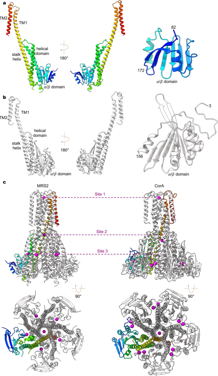
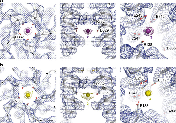
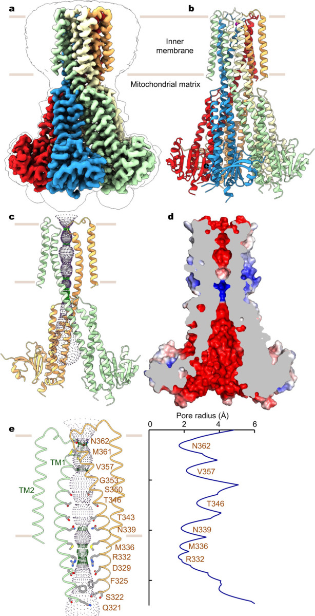
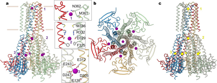
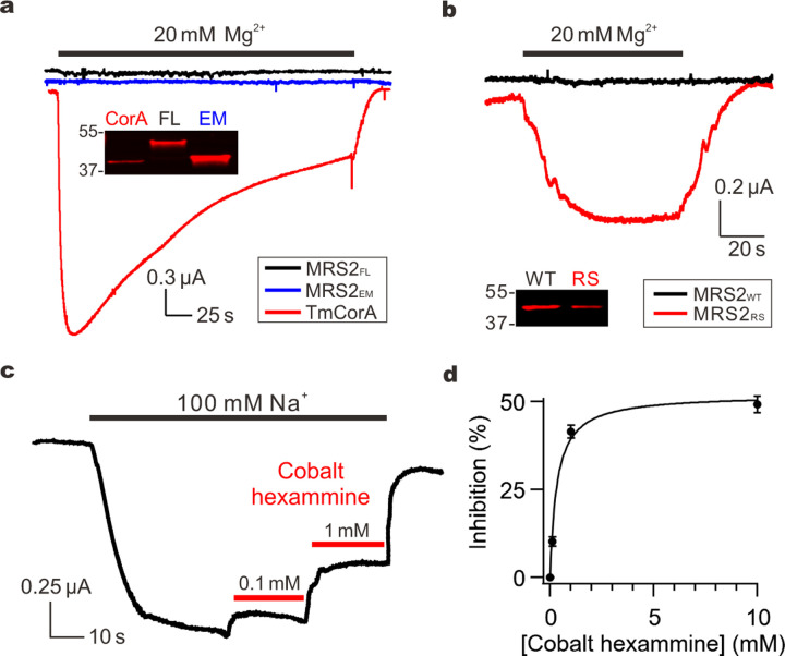
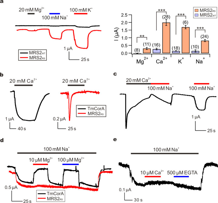
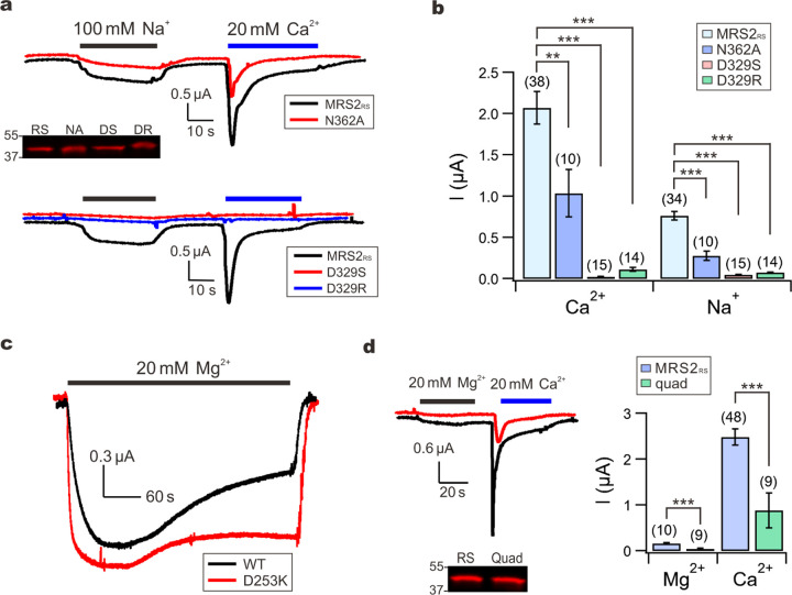
The human Mitochondrial RNA Splicing 2 protein (MRS2) has been implicated in Mg2+ transport across mitochondrial inner membranes, thus playing an important role in Mg2+ homeostasis critical for mitochondrial integrity and function. However, the molecular mechanisms underlying its fundamental channel properties such as ion selectivity and regulation remain unclear. Here, we present structural and functional investigation of MRS2. Cryo-electron microscopy structures in various ionic conditions reveal a pentameric channel architecture and the molecular basis of ion permeation and potential regulation mechanisms. Electrophysiological analyses demonstrate that MRS2 is a Ca2+-regulated, non-selective channel permeable to Mg2+, Ca2+, Na+ and K+, which contrasts with its prokaryotic ortholog, CorA, operating as a Mg2+-gated Mg2+ channel. Moreover, a conserved arginine ring within the pore of MRS2 functions to restrict cation movements, likely preventing the channel from collapsing the proton motive force that drives mitochondrial ATP synthesis. Together, our results provide a molecular framework for further understanding MRS2 in mitochondrial function and disease.


Conflict of interest statement

Competing Interests The authors declare no competing interests.

Figures

Extended Data Fig. 1 |
Extended Data Fig. 1 |. Cryo-EM data processing and validation.
a-c, Image processing and map validation of MRS2Mg (a), MRS2EDTA (b), and MRS2Ca (c). Representative micrographs and 2D classes are shown. The final 3D reconstructions are colored by local resolution. Also shown are Fourier shell correlations (FSC) and orientation distribution of particles used in the final reconstruction.
Extended Data Fig. 2 |
Extended Data Fig. 2 |. Cryo-EM density.
a, Ribbon representation of a single subunit of MRS2Mg and cryo-EM density. b, Segments of the final refined model of MRS2Mg and the corresponding cryo-EM densities.
Extended Data Fig. 3 |
Extended Data Fig. 3 |. Structural comparison of human MRS2 and TmCorA.
a-b, A single subunit of human MRS2 (a) and TmCorA (b, PDB ID: 4I0U). The N-terminal α/β domain is also highlighted for comparison. c, Divalent ion binding sites in human MRS2 (left panels) and TmCorA (right panels).
Extended Data Fig. 4 |
Extended Data Fig. 4 |. Sequence alignment of MRS2 channels.
Multiple protein sequences, including Homo sapiens MRS2 (hMRS2, NCBI sequence: NP_065713.1), Mus musculus MRS2 (mMRS2, NCBI sequence: NP_001013407.2), Rattus norvegicus MRS2 (rMRS2, NCBI sequence: NP_076491.1), Danio rerio MRS2 (zMRS2, NCBI sequence: XP_693621.5), Saccharomyces cerevisiae MRS2 (ScMRS2, NCBI sequence: NP_014979.1), Schizosaccharomyces pombe MRS2 (SpMRS2, NCBI sequence: NP_596358.1), Arabidopsis thaliana MRS2 (AtMRS2, NCBI sequence: AAM62917.1),and Thermotoga maritima CorA (TmCorA, NCBI sequence: WP_004081315.1). Secondary structure elements on the basis of hMRS2 are indicated above the sequences. Critical amino acids are highlighted.
Extended Data Fig. 5 |
Extended Data Fig. 5 |. Ion densities.
a-b, Cryo-EM densities in the three ion binding sites for Mg2+ (a) and Ca2+ (b). Mg2+ and Ca2+ are shown as magenta and yellow spheres, respectively.
Fig. 1 |
Fig. 1 |. Structure of human MRS2.
a, Cryo-EM reconstruction of human MRS2. Each subunit is uniquely colored and the contour indicates membrane boundaries. b, The overall structure of human MRS2. c, The central ion pathway, estimated using the HOLE program and represented as colored dots (pore radius: 1.15 Å < green < 2.3 Å < blue). Two opposite subunits are shown for clarity. d, Cutaway view of the channel surface, colored by surface electrostatic potential (red, −5 kT/e; white, neutral; blue, +5 kT/e). e, Details of the pore-lining residues and the dimension of the central pore.
Fig. 2 |
Fig. 2 |. Mg2+ and Ca2+ recognition.
a, Mg2+ ions (purple spheres) identified in the structure of MRS2 in the presence of Mg2+. Also shown are the details of the three Mg2+ ions (site 1 and 2) coordinated within the channel. Only two adjacent subunits are shown for clarity. b, Orthogonal view as in (a). c, Ca2+ ions (yellow spheres) in the MRS2Ca structure.
Fig. 3 |
Fig. 3 |. MRS2 currents.
a, Mg2+ transport by TmCorA or MRS2. The western blot image compares the expression of TmCorA, MRS2FL, and MRSEM, all tagged with a C-terminal 1D4 sequence and detected with an anti-1D4 antibody. b, MRS2 Mg2+ currents induced by the R332S mutation. MRS2RS exhibits ~60% expression level of MRS2WT. c, Inhibition of MRS2 activity by cobalt hexammine. Na+ currents were presented here, but similar inhibition was obtained when recording Mg2+ currents. d, Dose response of cobalt hexammine inhibition of MRS2 Na+ currents. Data points were fit with a standard saturation binding equation (black curve). Maximal inhibition achieved is ~50%, comparable with that obtained with TmCorA.
Fig. 4 |
Fig. 4 |. Ion permeation properties.
a, Monovalent and divalent cation currents through MRS2WT or MRS2RS. Peak-level amplitudes of Ca2+ currents were presented in the bar chart. **P < 0.01; ***p < 0.001. b, Ca2+ currents conducted by TmCorA or MRS2RS. c, Recovery of MRS2RS from inactivation. d, The effect of Mg2+ on Na+ currents. e, Lack of Ca2+ inhibition of MRS2 Na+ currents. 500 µM of EGTA was added later in the recording to test if the trace amount of Ca2+ in the NaCl buffer can inhibit Na+ currents.
Fig. 5 |
Fig. 5 |. Divalent cation-binding sites.
a-b, Functional impacts of site 1 or site 2 mutations on MRS2RS. The Western-blot image shows that these mutations do not affect MRS2RS expression. RS: MRS2RS. NA: N362A; DS: D329S; DR: D329R. c, Mg2+ inactivation of TmCorA. d, The effect of site 3 mutations on MRS2RS function (quad: E138A-E243A-D247A-E312A mutation). Western blot shows that these mutations do not reduce surface expression.

References

    1. de Baaij J. H., Hoenderop J. G. & Bindels R. J. Magnesium in man: implications for health and disease. Physiol Rev 95, 1–46 (2015). https://doi.org: 10.1152/physrev.00012.2014 - DOI - PubMed
    1. Jin F., Huang Y. & Hattori M. Recent Advances in the Structural Biology of Mg(2+) Channels and Transporters. J Mol Biol 434, 167729 (2022). https://doi.org: 10.1016/j.jmb.2022.167729 - DOI - PubMed
    1. Quamme G. A. Molecular identification of ancient and modern mammalian magnesium transporters. Am J Physiol Cell Physiol 298, C407–429 (2010). https://doi.org: 10.1152/ajpcell.00124.2009 - DOI - PubMed
    1. Wiesenberger G., Waldherr M. & Schweyen R. J. The nuclear gene MRS2 is essential for the excision of group II introns from yeast mitochondrial transcripts in vivo. J Biol Chem 267, 6963–6969 (1992). - PubMed
    1. Zsurka G., Gregan J. & Schweyen R. J. The human mitochondrial Mrs2 protein functionally substitutes for its yeast homologue, a candidate magnesium transporter. Genomics 72, 158–168 (2001). https://doi.org: 10.1006/geno.2000.6407 - DOI - PubMed

Publication types